The calf is an area at the back of the leg. It brings together two bones and three muscles, which are essential for the movements of the knee, ankle and foot. To learn more about this anatomical part located in the popliteal fossa, read on.
You will find all the information related to the biomechanics of the calf and knee and a detail of the most common gastrocnemius injuries caused by sports activities and other ailments. You can also take a look at treatments and products to help relieve soleus and calf pain.
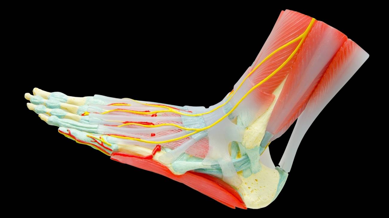
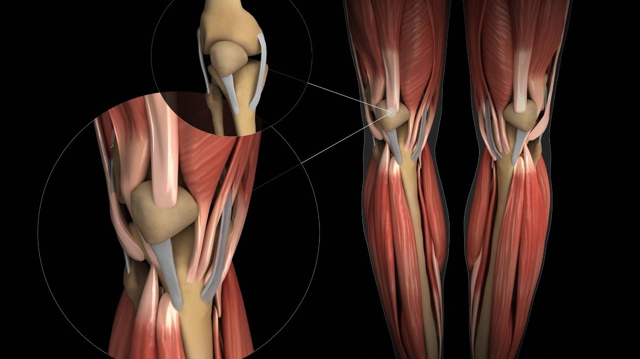
Calf parts and anatomy
The anatomical parts that make up the calf are:
Bones and joints
The bones of the calf are as follows:
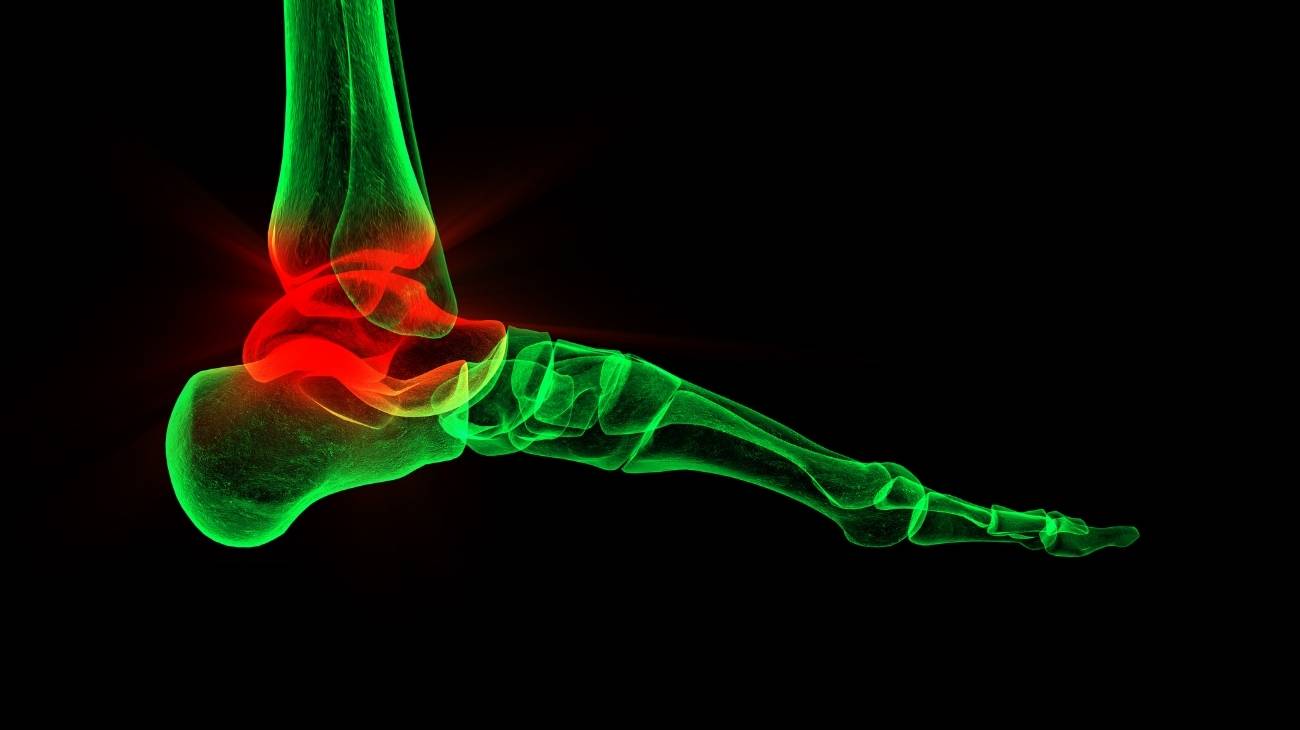
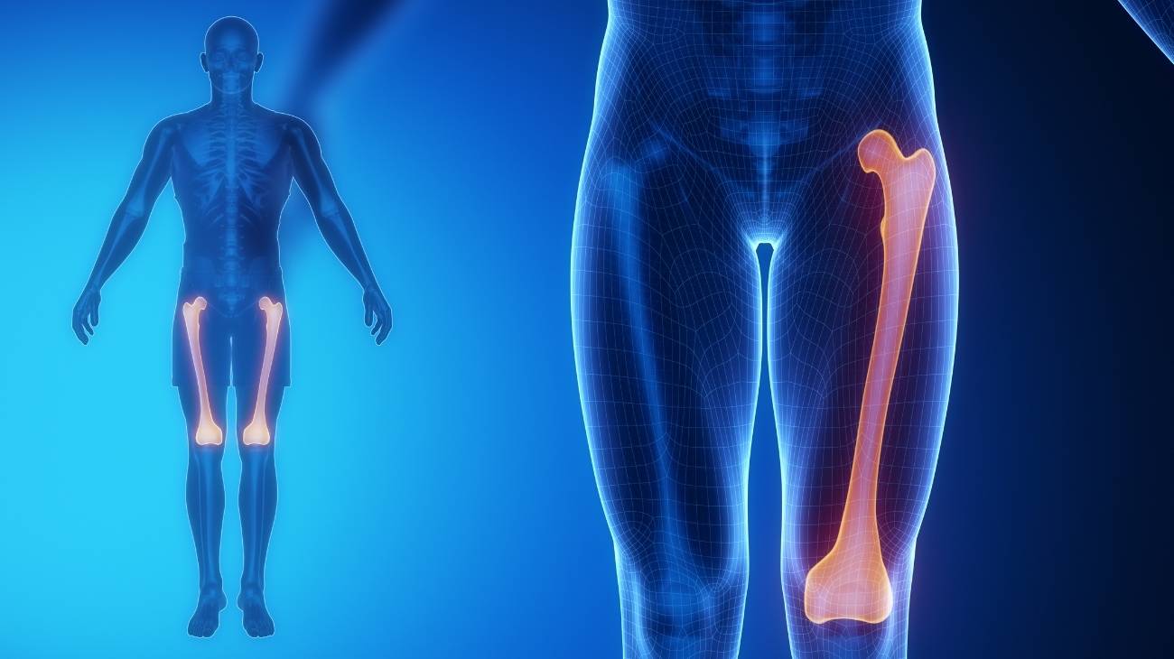
- Tibia: This elongated, triangular-shaped bone runs along the anterior part of the calf. It is joined in the upper area, by means of the glenoid cavities, with the femoral condyles, giving rise to the knee joint. On the lower plane, it is connected to the upper aspect of the talus (forming the ankle).
- Fibula: Also known as fibula, it runs along the posterior aspect of the calf, parallel to the tibia. The lower part of the fibula has a malleolus that is joined to the talus and calcaneus by ligaments.
Take a look at the joints of the calf:
- Proximal or superior tibioperoneal: It is responsible for making small movements between the head of the fibula and the lateral condyle of the tibia.
- Distal tibioperoneal: This is the only syndesmosis-type joint in the leg. It is a membrane-like joint located between the tibia and fibula, thus generating the interosseous ligament.
- Tibioperoneoastotalar: Also known as the tibiotarsal joint. It is a synovial joint and allows movement of the lower part of the tibia and fibula with the talus.

Muscles
Learn about the muscle structure of the calves:
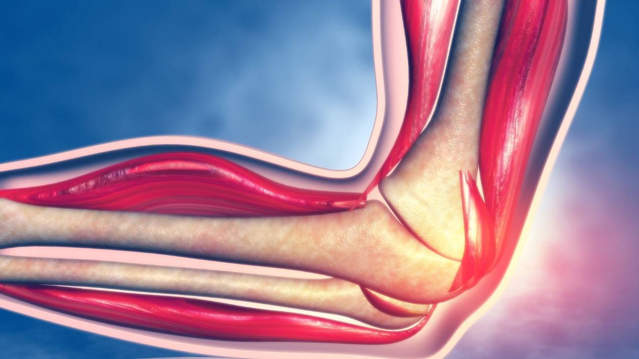
- Gastrocnemius or calf: Its function is plantar flexion of the foot. It is a muscle that originates in the femoral condyles and separates into two, always with a route above the soleus. It inserts into the Achilles tendon together with the soleus tendon, so it can be divided into three zones: medial head, lateral head and lower portion.
- Soleus: It is also responsible for plantar flexion. Its origin is multipeniform, it is produced in the membrane of the tibia and fibula, in the simular head and in the medial border of the tibia. It inserts on the soleus tendon to form the Achilles tendon, together with the calf tendon, and on the posterior surface of the calcaneal bone.

Ligaments
The connective tissues that belong to the calves are:
- Anterior of the fibular head: It is responsible for joining the lateral condyle of the tibia to the head of the fibula, by means of three wide bands.
- Posterior of the fibular head: Unlike the anterior connective tissue, this ligament is a single band that runs from the head of the fibula to the lateral condyle of the tibia, in the posterior part.
- Interosseous: A tissue located between the tibia and fibula, which works with the distal tibioperoneal joint.
- Anterior peroneoastotalar: originates from the lateral malleolus of the fibula to the neck of the talus.
- Posterior peroneoastotalar: The distal fibular end gives rise to this ligament, which inserts into the talus through its lateral tubercle.
- Peroneocalcaneal: This ligament runs from the apex of the lateral malleolus of the fibula to a tubercle of the calcaneus.

Best products for leg and calf pain relief
Bestseller
-

2 Calf Compression Sleeve (Black/Gray)
$19.95 -
2 Calf Compression Sleeve (Green/Navy)
$19.95 -
2 Calf Compression Sleeve (Pink/Bordeaux)
$19.95 -
Acupressure Mat and Pillow (Black/Gray)
$49.95 -
Acupressure Mat and Pillow (Green/Navy)
$49.95 -
Acupressure Mat and Pillow (Pink/Bordeaux)
$49.95 -
Acupressure Pillow (Black/Gray)
$29.46 -
Acupressure Pillow (Green/Navy)
$29.46 -
Acupressure Pillow (Pink/Bordeaux)
$29.46 -
Electric Muscle Massager Gun
$177.03 -

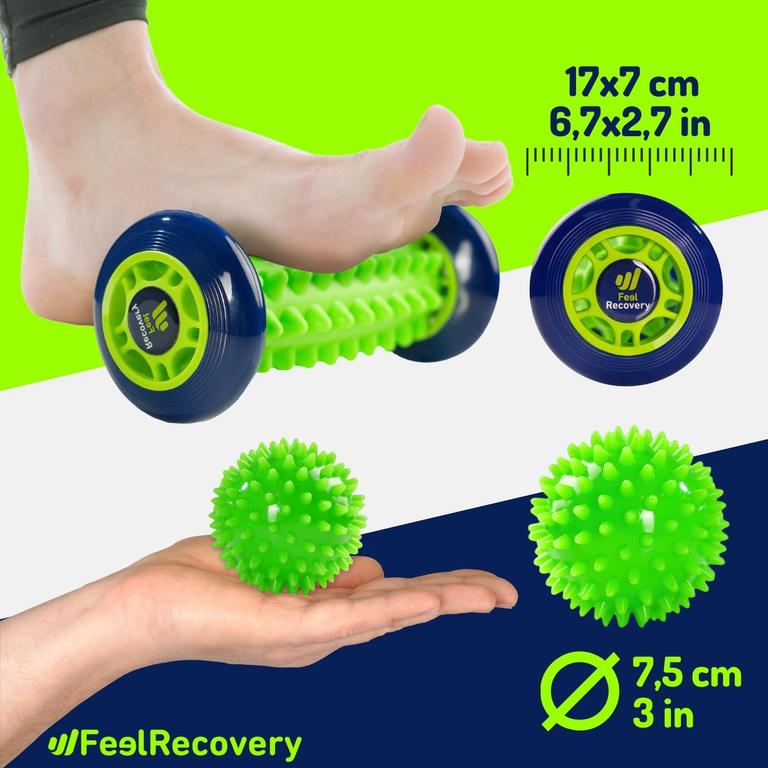
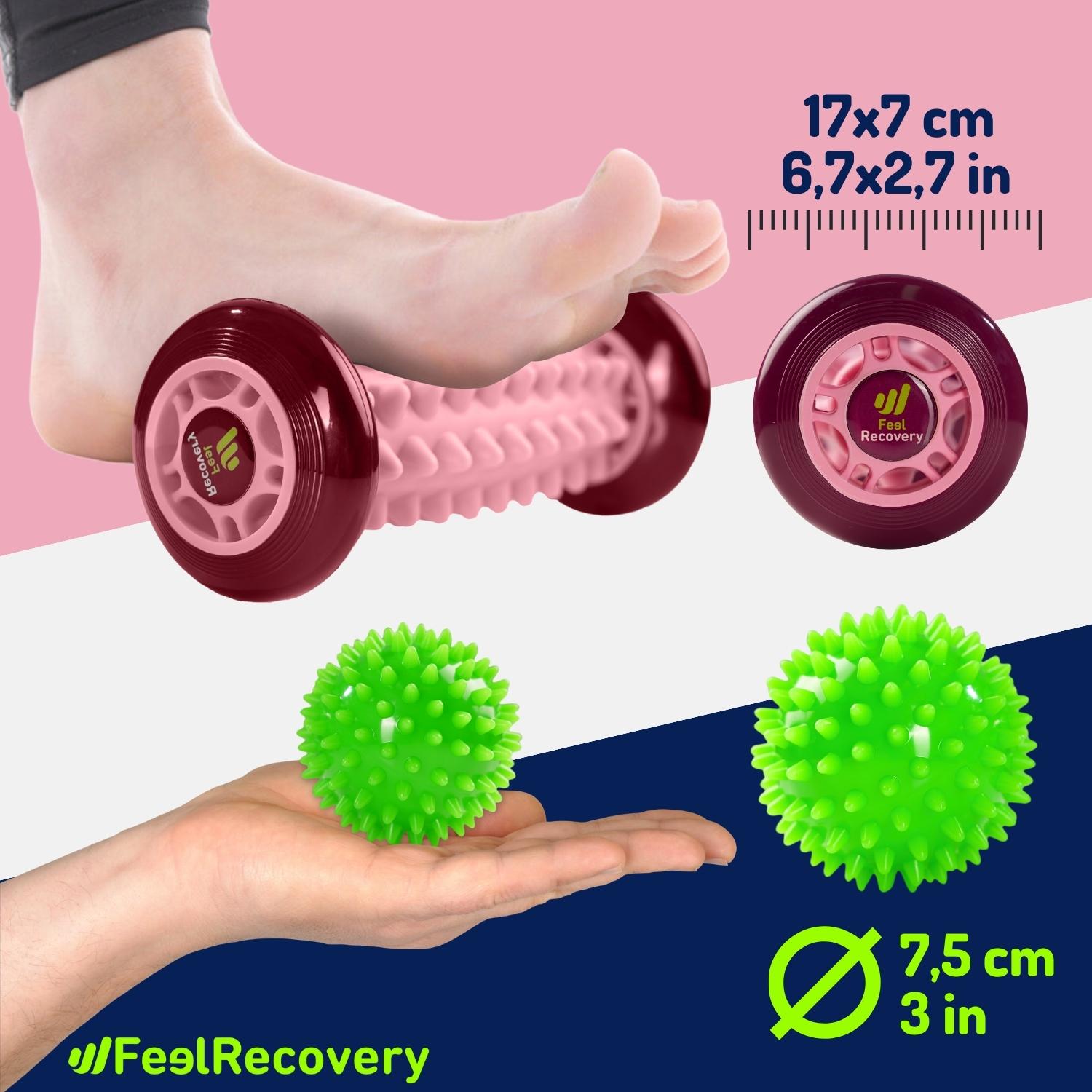
Foot Massage Roller for Plantar Fasciitis (Black)
$19.95 -
Foot Massage Roller for Plantar Fasciitis (Green)
$19.95 -
Foot Massage Roller for Plantar Fasciitis (Pink)
$19.95 -
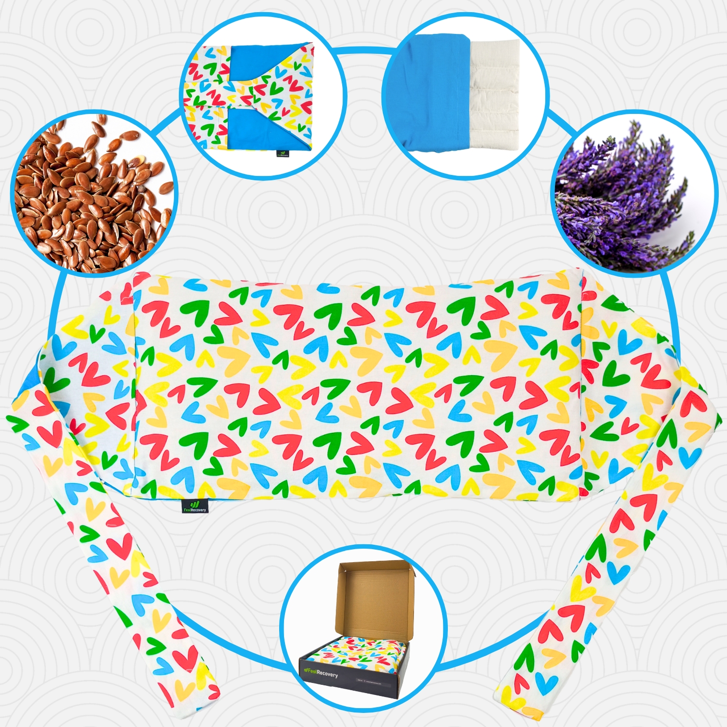
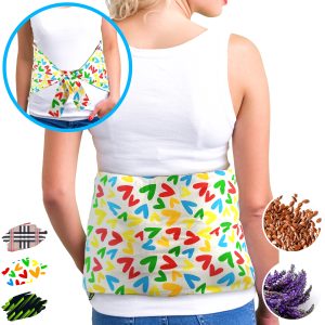
Heating Pad for Microwave Classic Bottle Shaped (Hearts)
$19.95 -
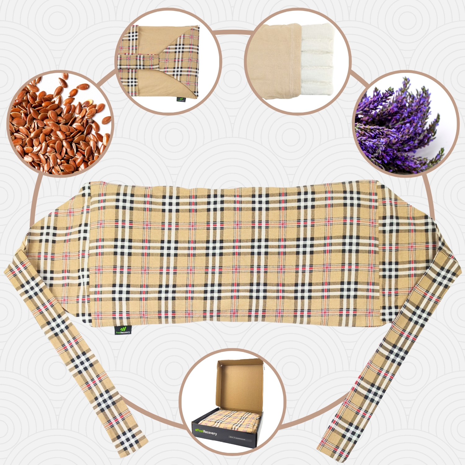
Heating Pad for Microwave Classic Bottle Shaped (Oxford)
$19.95 -
Heating Pad for Microwave Classic Bottle Shaped (Sport)
$19.95 -
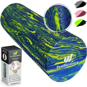
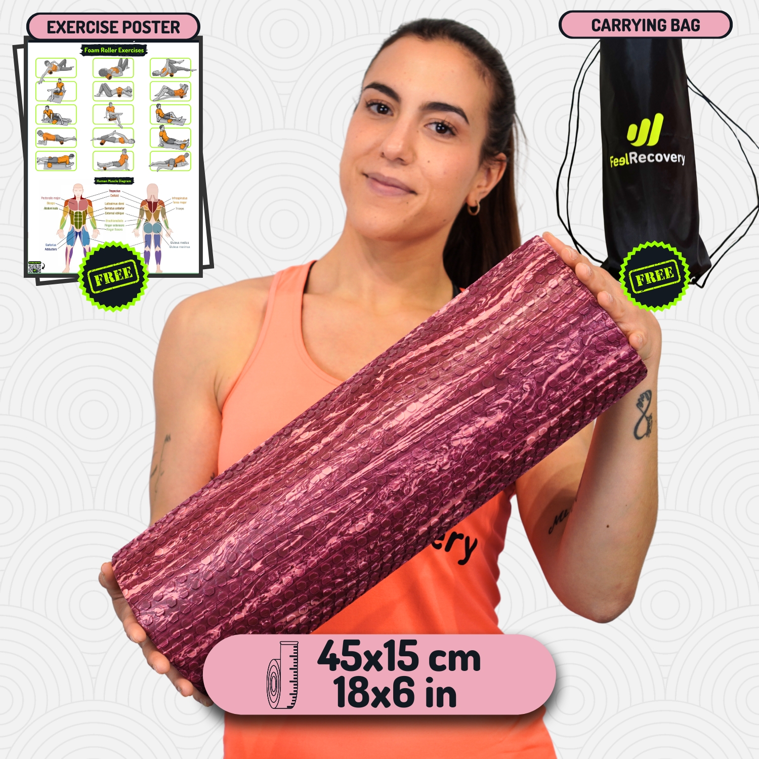
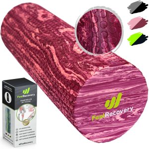
High Density Foam Roller for Muscle (Black/Gray)
$29.95 -
High Density Foam Roller for Muscle (Green/Navy)
$29.95 -
High Density Foam Roller for Muscle (Pink/Bordeaux)
$29.95 -
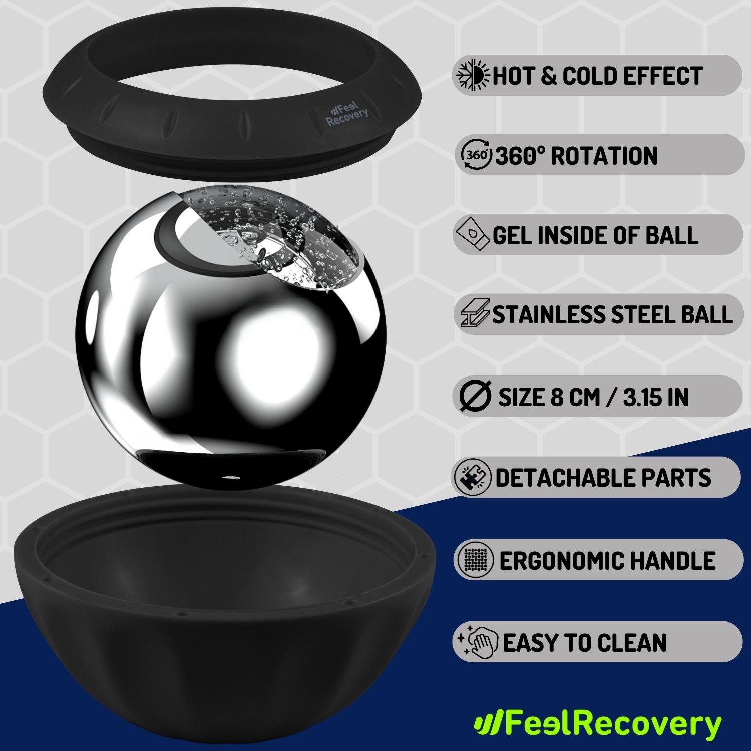
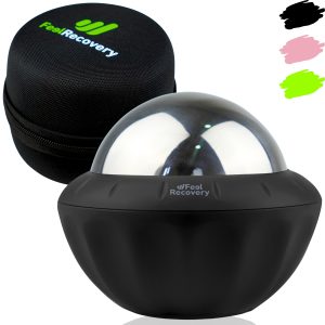
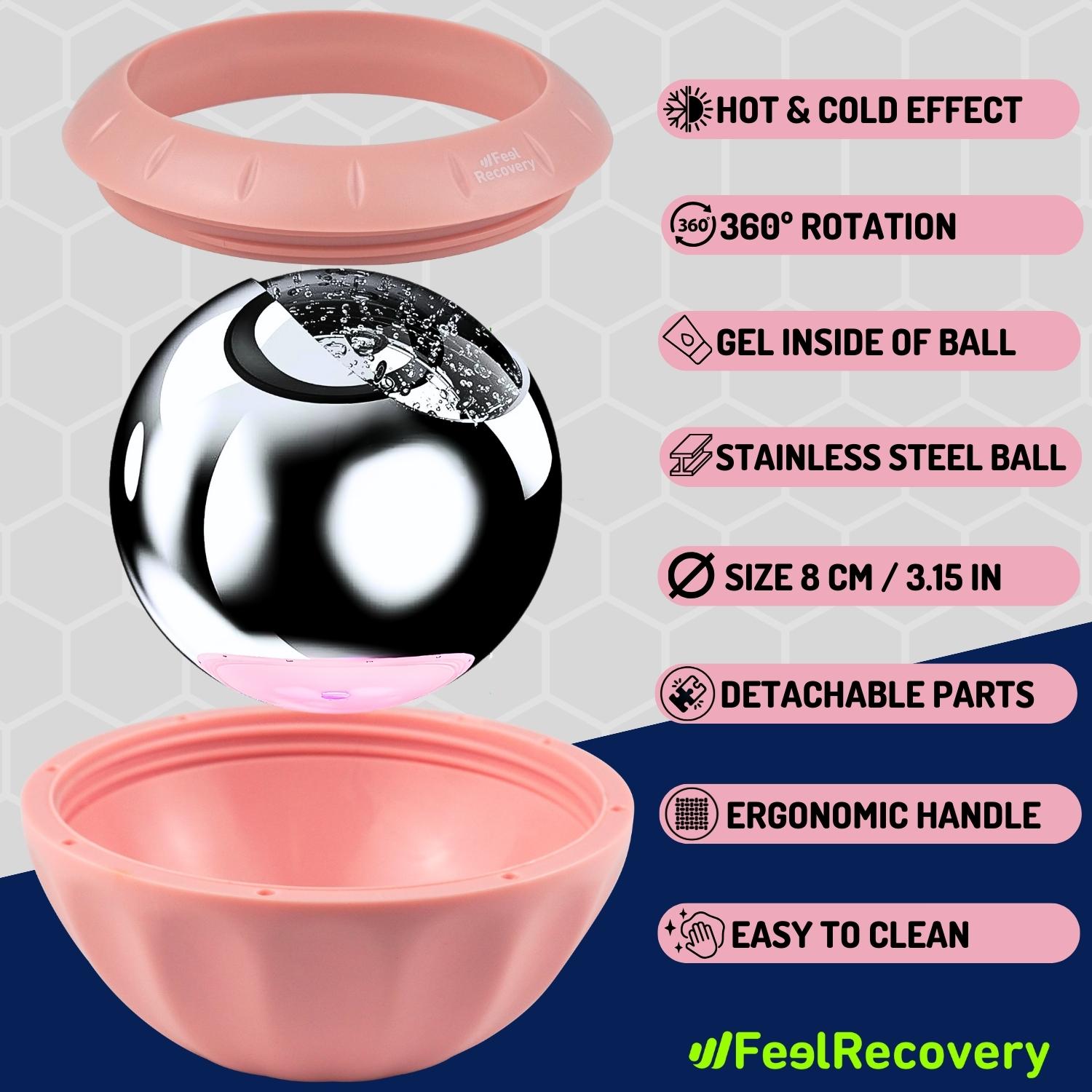
Ice Massage Roller Ball (Black)
$39.95 -
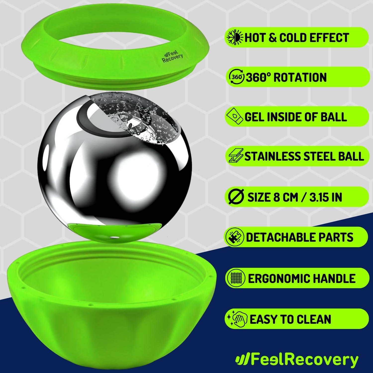
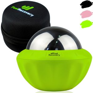
Ice Massage Roller Ball (Green)
$39.95 -
Ice Massage Roller Ball (Pink)
$39.95 -
Microwave Heating Pad for Back Pain Relief (Extra Large) (Hearts)
$29.95 -
Microwave Heating Pad for Back Pain Relief (Extra Large) (Oxford)
$29.95 -
Microwave Heating Pad for Back Pain Relief (Extra Large) (Sport)
$29.95 -
Microwaveable Heating Pad for Pain Relief (Hearts)
$19.95 -
Microwaveable Heating Pad for Pain Relief (Oxford)
$19.95 -
Microwaveable Heating Pad for Pain Relief (Sport)
$19.95 -
Mini Electric Massager Gun
$58.97 -
Pack 2 In 1 Foam Roller High + Soft Density (Black/Gray)
$29.95 -
Pack 2 In 1 Foam Roller High + Soft Density (Green/Navy)
$29.95 -
Pack 2 In 1 Foam Roller High + Soft Density (Pink/Bordeaux)
$29.95 -
Pack 2 Peanut Massage Rollers
$19.95 -
Pack 3 Spiky Massage Balls
$19.95 -
Pack Lacrosse Massage Balls & Peanut Roller
$29.95 -
Reusable Extra Large Ice Packs for Back & Legs Pain
$29.95 -
Reusable Gel Ice Packs for Knees with Compression Band
$19.95 -
Reusable Gel Ice Packs with Compression Band for Sports Injury
$19.95 -
Soft Density Foam Roller for Recovery (Black)
$29.95 -
Soft Density Foam Roller for Recovery (Green)
$29.95 -
Soft Density Foam Roller for Recovery (Pink)
$29.95 -
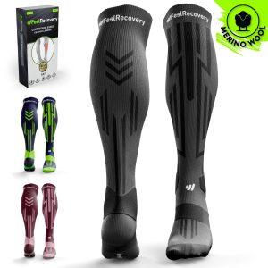
Sport Compression Socks (1 Pair) (Black/Gray)
$19.95 -
Sport Compression Socks (1 Pair) (Green/Navy)
$19.95 -
Sport Compression Socks (1 Pair) (Pink/Bordeaux)
$19.95 -
Trigger Point Massage Stick (Black)
$14.95 -
Trigger Point Massage Stick (Green)
$14.95 -
Trigger Point Massage Stick (Pink)
$14.95 -
Vibrating Foam Roller
$35.36 -
Vibrating Massager Ball
$35.36


Biomechanics of the leg (calf and knee)
The biomechanical movements performed by the calf and knees are
- Extension: This action consists of keeping the leg at rest. That is, it is to place the tibia and fibula in a straight line in relation to the axis of the femur, so the amplitude is 0°.
- Flexion: This is the opposite movement to extension and is performed with an opening of 130°, although it can reach 170° with the help of the hand. It involves bringing the heel to the buttock, causing the cruciate ligaments, menisci and quadriceps to offer resistance.
- External rotation: If the tibia is considered as the axis, this biomechanical action is to bring the distal part of the foot towards the side of the fibula. Its maximum amplitude can reach 90° with a lot of training, as is the case with ballet dancers.
- Internal rotation: It is possible to biomechanically rotate the foot towards the internal axis of the tibia, thanks to the action of the calf, soleus, lateral ligaments and tendons, which prevent the displacement of the tibia with respect to the fibula.
- Plantar flexion: This movement is produced by the action of the gastrocnemius and soleus muscles. It involves placing the tibia at a maximum angle of 90° to the dorsal part of the foot.
- Dorsal flexion: This consists of bringing the toes towards the tibia, forming an angle that does not exceed 30° of opening. This biomechanical movement is carried out by the action of different elements, including the gastrocnemius and soleus.
Most common calf injuries
There are different types of calf injuries that can occur. We will show you below the most common ones.
Types of calf injuries
The most common calf injury is a calf strain
- Muscle contractures in the calf muscles: Involuntary tensions caused in the soleus and gastrocnemius are caused by the accumulation of metabolites in the muscle fibres. This is caused by the inability of the blood to exchange oxygen and nutrients. This injury is caused by a variety of factors, the most common being muscle overload and trauma.
- Legs and calf cramps: In general terms, muscle cramps are defined as a contraction of the muscles that prevents them from relaxing and, at the same time, causes great temporary pain. Specifically, leg or calf cramps are those spasms that appear suddenly in the muscles connected to these limbs and appear as a kind of twinge.
Soleus and calf sports injuries
Sports also cause different calf injuries in athletes, take a look at the most common ones
- Calf injuries in soccer: Hamstring and calf contractures are the most common injuries suffered by these athletes. It is also possible to find calf tears caused by sudden movements, jumps and demanding actions.
- Calf injuries in running: In this sport, athletes also suffer from contractures and tears in the soleus and gastrocnemius muscles. This is due to the constant muscular strain, the lack of warm-up and adequate stretching.
Diseases and ailments in the calf muscles

Diseases that can occur in twins, in addition to those mentioned above, usually include
Fibromyalgia
This disease causes a deterioration of the muscles and ligaments located in the calf. It is a chronic pain that causes sleep disturbances, headaches, changes between constipation and diarrhoea and fatigue. Its causes are unknown.
Duchenne syndrome
Duchenne syndrome is an inherited disorder that causes progressive weakness in the calf and other muscles. Patients with this condition suffer from sudden falls, difficulty walking and enlarged calves, among other symptoms.
Progressive muscular dystrophy
The alterations that occur in muscle mass due to this chronic degenerative disease cause a considerable decrease in proteins to maintain the calves, soleus and other tissues in good condition. There are different types of muscular dystrophy, but the most common is progressive muscular dystrophy and Duchenne muscular dystrophy.
Cramps
This condition occurs in the calf muscles, usually when waking up from a night's rest or when falling asleep due to lack of physical activity or too much physical activity. This is caused by a lack of potassium and magnesium in the body.
How can we relieve calf pain through complementary and non-invasive therapies?
The different treatments that help to relieve calf pain are listed below:
Heat and cold therapy
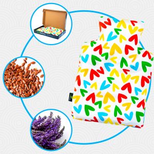
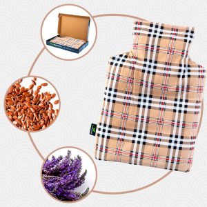
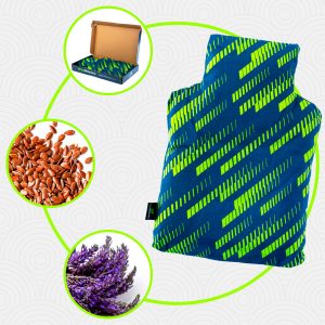
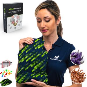
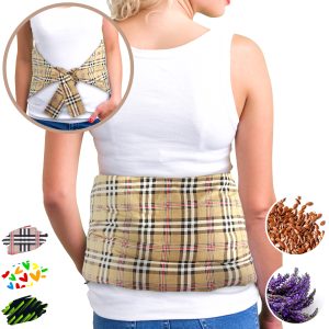
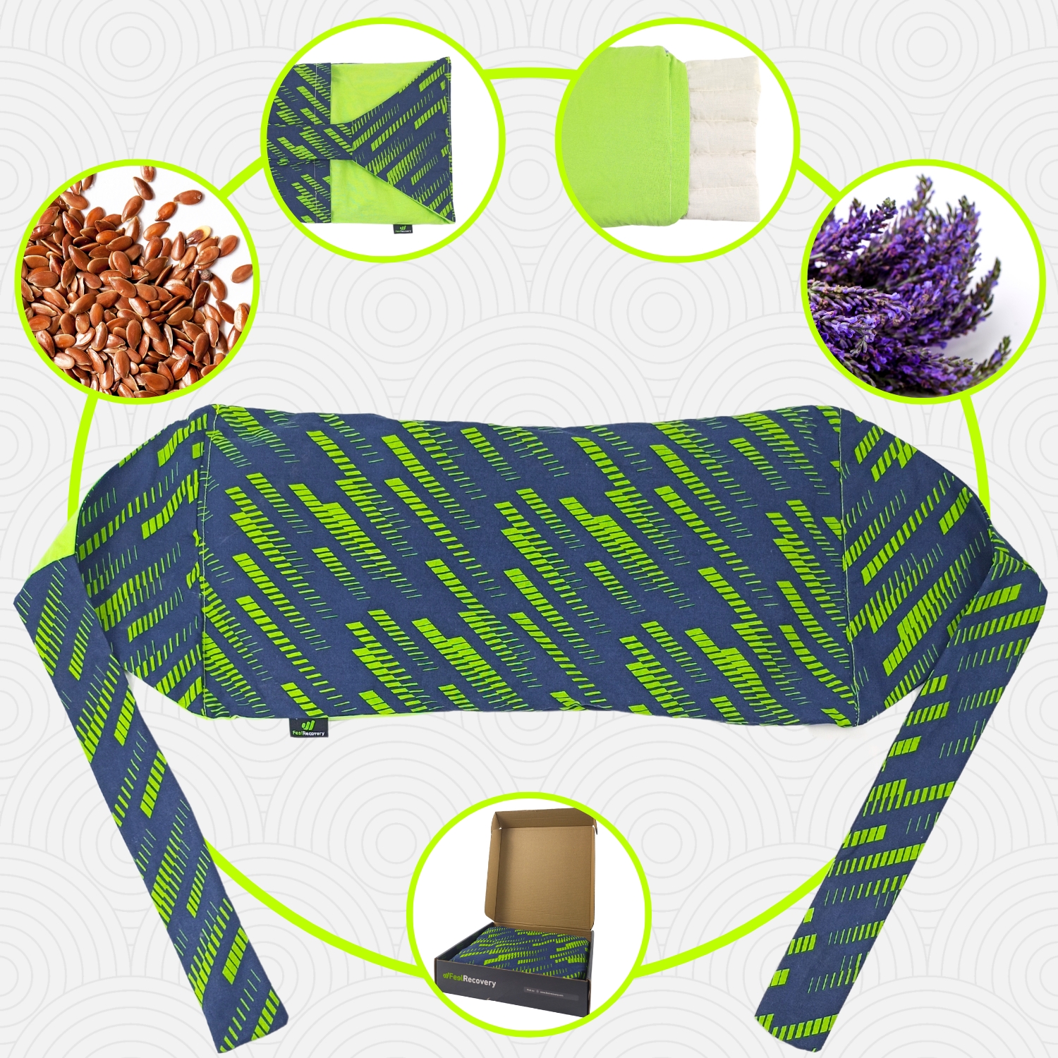
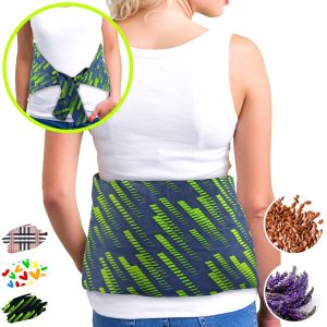
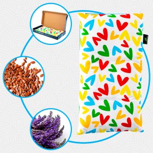
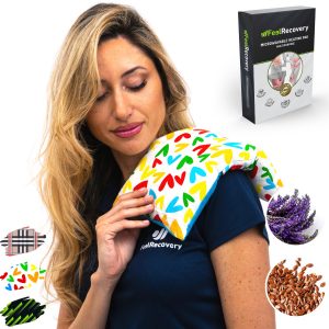
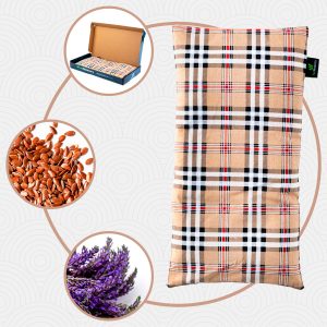
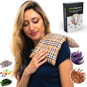
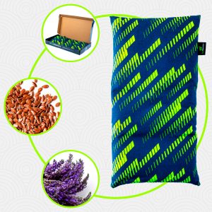
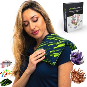
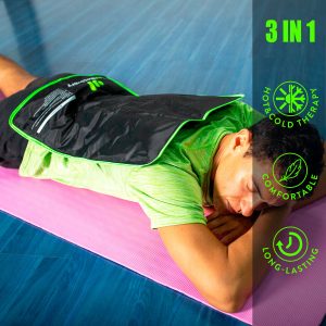
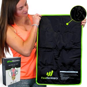
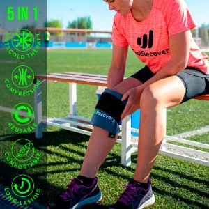
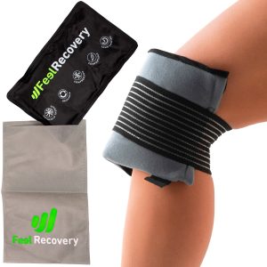
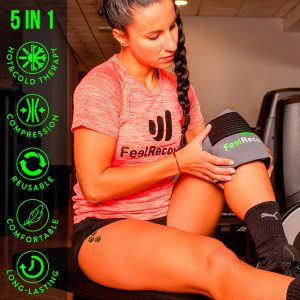
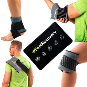
The application of heat and cold in a single session lasting no longer than 20 minutes will help to reduce calf pain. This will be generated by the anti-inflammatory and vessel dilating properties of both temperatures. Cold gel packs with compression strap can be used to improve recovery time. It is advisable to consult a doctor before applying this non-invasive therapy.
Compression therapy
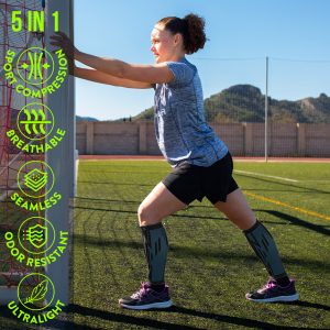
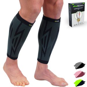
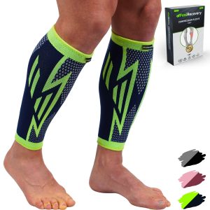
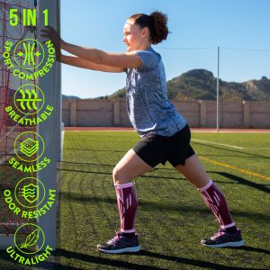
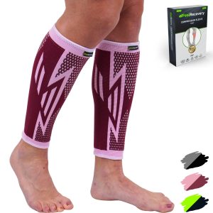
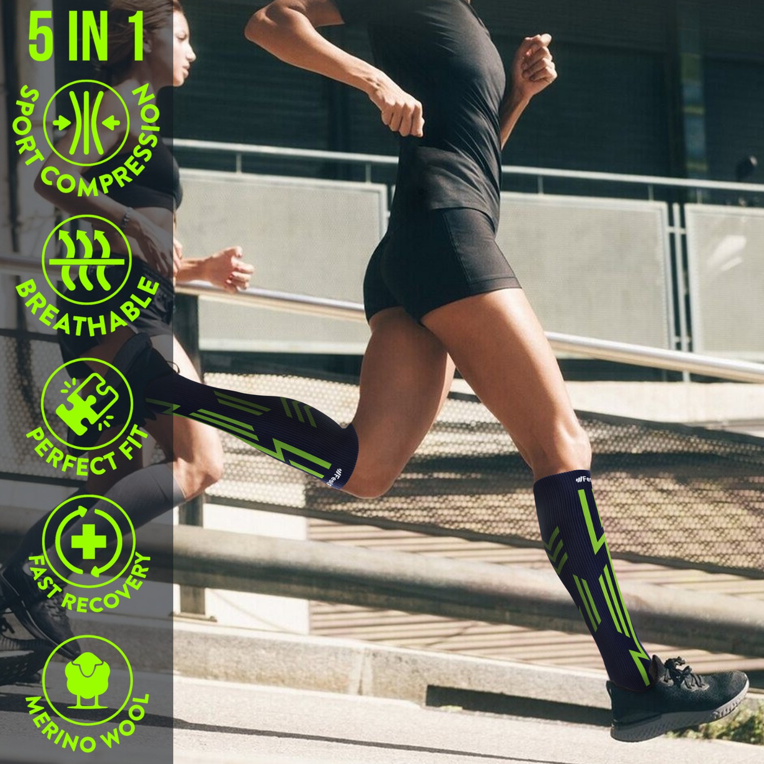
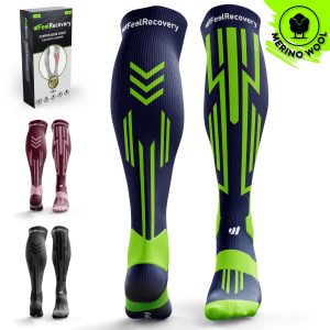
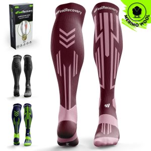
This type of therapy consists of holding the calf still so that the muscles can rest and eliminate the molecules with toxic substances found in the fibres. For this purpose, it is important to use elastic bandages, compression calf sleeves, sports knee braces, compression ankle braces and compression sports socks, among other products. The advantage of these items is that they will fit the body in the right way and with the right pressure.
Massage therapy
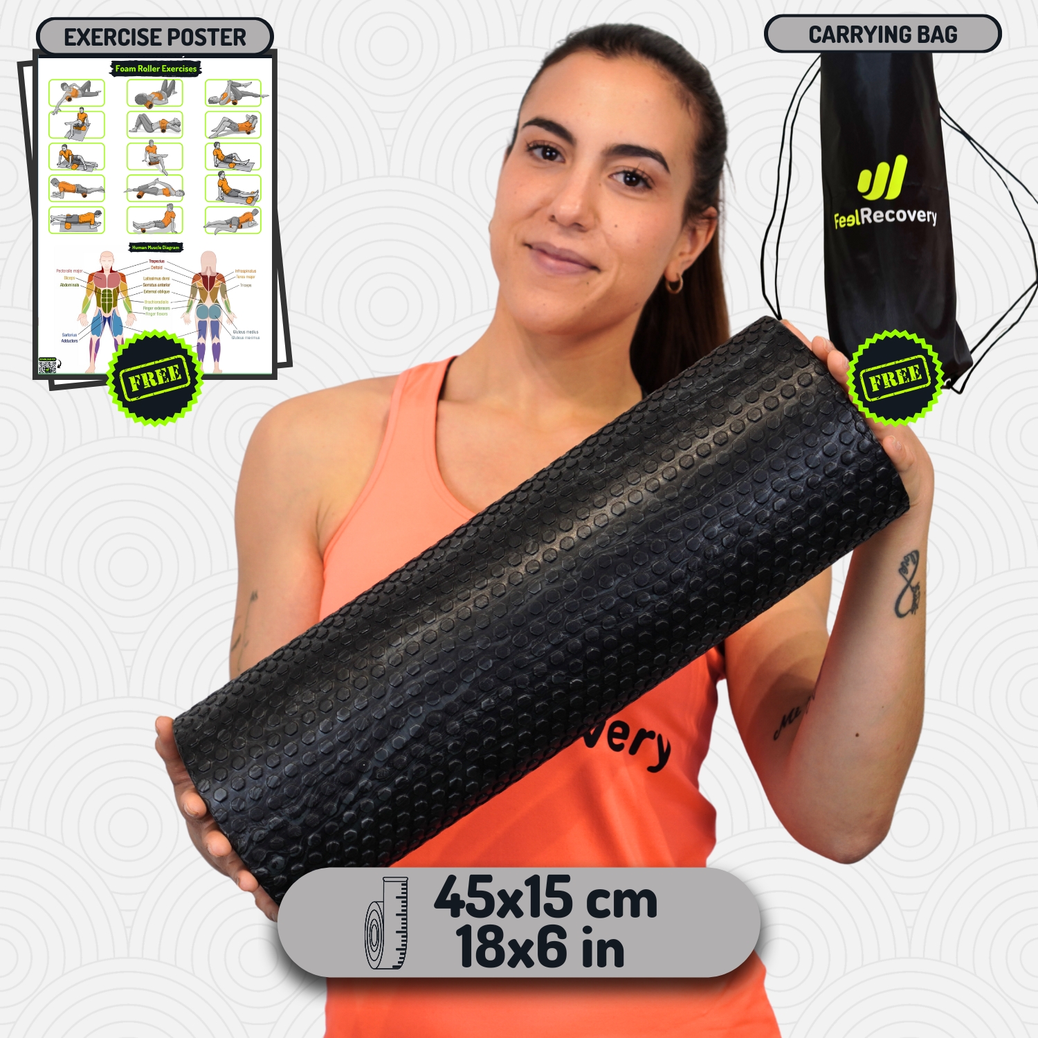
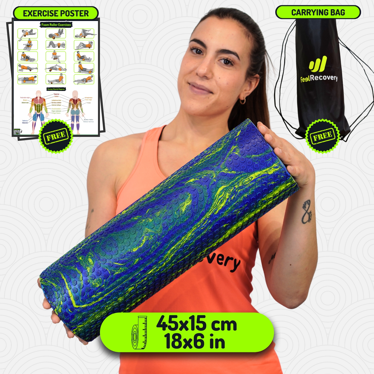
This therapeutic alternative consists of massaging the soleus and gastrocnemius muscles to stimulate blood circulation and decompress involuntary contractures. This can be done in different ways, but it is currently recommended to use non-invasive products that achieve effective results in the short term. For this, electric massage guns, vibrating balls and massage cushions can be used.
Acupressure therapy
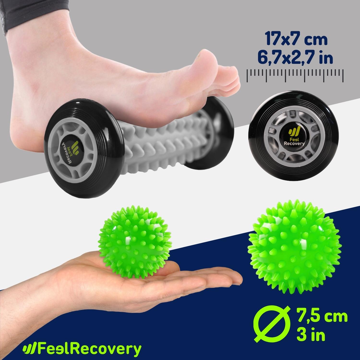
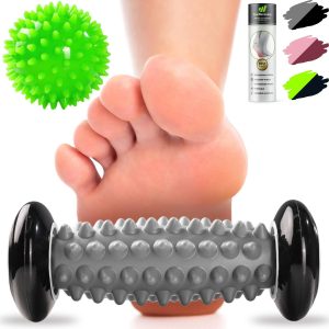
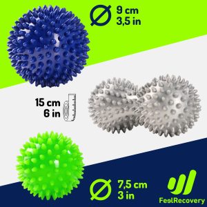
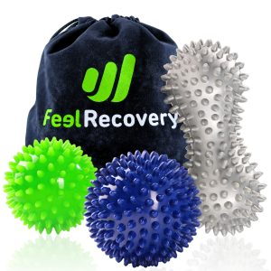
This is a very effective complementary therapy to reduce the symptoms of calf pain. To do this, it is necessary to choose between various products that will help to stimulate the blood flow in this part of the body to eliminate lactic acid. You will be able to use, among other items, massage hooks, massage balls and manual foot massagers with rollers. Don't forget that these products will help you save money, as you can also visit a professional therapist.
Thermotherapy
Thermotherapy improves the symptoms of pain and inflammation in the calf and soleus caused by bruises and other injuries. This treatment involves applying the desired temperature, in a controlled manner, to the affected area using microwavable heat packs or hot and cold gel packs. This will improve the dilation of the capillary walls to help blood flow.
Cryotherapy
Inflammations in the gastrocnemius and calf can be treated by cryotherapy. This non-invasive treatment involves using the benefits of cold to produce a decrease in pain and improve vasodilation. Different techniques can be implemented, but the most commonly used at present are hot/cold packs with special gels . In some cases the doctor may recommend cold chambers.
Electrical muscle stimulation (EMS)
Muscle electrostimulation, or EMS, is a therapy that consists of stimulating muscle contractions through the use of electricity, so as to achieve an effect of activity and hypertrophy as in the gym, but without the need to go to any sports center. This means that you can put your muscles to work without leaving home.
Electrotherapy
This is a technique that seeks relief from pain and some physical ailments through the application of electrical and electromagnetic energy, among other variants, through the skin with the use of conductive pads called electrodes. It is a very safe type of therapy and must be applied by a physical therapist specialized in the manipulation of electricity to treat some kinds of ailments.
Myofascial release therapy
Also known as myofascial induction, this therapy consists of the application of manual massage to treat the shortening and tension generated in the myofascial tissue that connects the muscles to the bones and nerves. For this purpose, various massage techniques are used that focus on the so-called trigger points.
Percussion Massage Therapy
Vibration or percussion massages are precise, rhythmic and energetic strokes on the body to achieve relief from some annoying symptoms when muscle fibers are tightened, often by a high workload on them and that has left trigger points in the muscle fibers.
R.I.C.E Therapy
The R.I.C.E. therapy is the first and simplest of the treatment protocols for minor injuries. It appears in the sports field to deal with accidents involving acute injuries. For many years, it has been considered the most suitable for its speed and results.
Trigger points therapy
Myofascial pain points or trigger points are knots that are created in the deeper muscle tissues, causing intense pain. The pain does not always manifest itself right in the area where the point develops, but rather this pain is referred to nearby areas that seemingly do not appear to be related. In fact, it is estimated that more than 80% of the pain they cause manifests in other parts of the body.
Other effective alternative therapies
It is also possible to choose alternative, non-invasive therapies to help improve the symptoms of calf pain.
- Natural remedies using plants: This therapy can be applied by means of tesinas or footbaths to obtain the anti-inflammatory and relaxation benefits of natural plants. Linden, chamomile and willow bark, among other plants, can be used.
- Acupuncture: This is an oriental technique widely used to obtain mental balance and reduce the patient's stress in the face of pain symptoms in the calves. It is used by means of special needles in strategic places of the body.
- Kinesiotherapy: Pain in the calf and soleus can be combated by means of special exercises performed by professionals on the legs and ankles. This improves joint range of motion and combats contractures and sprains.
- Aromatherapy: This technique aims to reduce the stress caused by an injury to the calf muscles. Aromas of citrus fruits, ginger and mint can be applied in oils or liquids for diffusers.
- Osteopathy: This is not a scientifically proven therapy, but it can be used to treat calf pain. It is a set of exercises that stimulate the body's natural recovery.
References
- Bordoni, B., & Varacallo, M. (2018). Anatomy, bony pelvis and lower limb, gastrocnemius muscle. https://europepmc.org/article/NBK/nbk532946
- Maganaris, C. N., Baltzopoulos, V., & Sargeant, A. J. (2006). Human calf muscle responses during repeated isometric plantarflexions. Journal of biomechanics, 39(7), 1249-1255. https://www.sciencedirect.com/science/article/abs/pii/S0021929005001508
- Bryan Dixon, J. (2009). Gastrocnemius vs. soleus strain: how to differentiate and deal with calf muscle injuries. Current reviews in musculoskeletal medicine, 2(2), 74-77. https://link.springer.com/article/10.1007/s12178-009-9045-8
- Green, B., & Pizzari, T. (2017). Calf muscle strain injuries in sport: a systematic review of risk factors for injury. British journal of sports medicine, 51(16), 1189-1194. https://bjsm.bmj.com/content/51/16/1189?hootPostID=8dbac92262359e5c3a3438f4e11668c6
- Prakash, A., Entwisle, T., Schneider, M., Brukner, P., & Connell, D. (2018). Connective tissue injury in calf muscle tears and return to play: MRI correlation. British journal of sports medicine, 52(14), 929-933. https://bjsm.bmj.com/content/52/14/929.abstract
- Fields, K. B., & Rigby, M. D. (2016). Muscular calf injuries in runners. Current sports medicine reports, 15(5), 320-324. https://journals.lww.com/acsm-csmr/fulltext/2016/09000/Muscular_Calf_Injuries_in_Runners.9.aspx
- Fahlström, M., Jonsson, P., Lorentzon, R., & Alfredson, H. (2003). Chronic Achilles tendon pain treated with eccentric calf-muscle training. Knee surgery, sports traumatology, arthroscopy, 11(5), 327-333. https://link.springer.com/article/10.1007/s00167-003-0418-z
- Radford, J. A., Landorf, K. B., Buchbinder, R., & Cook, C. (2007). Effectiveness of calf muscle stretching for the short-term treatment of plantar heel pain: a randomised trial. BMC musculoskeletal disorders, 8(1), 1-8. https://bmcmusculoskeletdisord.biomedcentral.com/articles/10.1186/1471-2474-8-36
- Jonsson, P., Alfredson, H., Sunding, K., Fahlström, M., & Cook, J. (2008). New regimen for eccentric calf-muscle training in patients with chronic insertional Achilles tendinopathy: results of a pilot study. British journal of sports medicine, 42(9), 746-749. https://bjsm.bmj.com/content/42/9/746.short
- Alfredson, H., Pietilä, T., Jonsson, P., & Lorentzon, R. (1998). Heavy-load eccentric calf muscle training for the treatment of chronic Achilles tendinosis. The American journal of sports medicine, 26(3), 360-366. https://journals.sagepub.com/doi/abs/10.1177/03635465980260030301