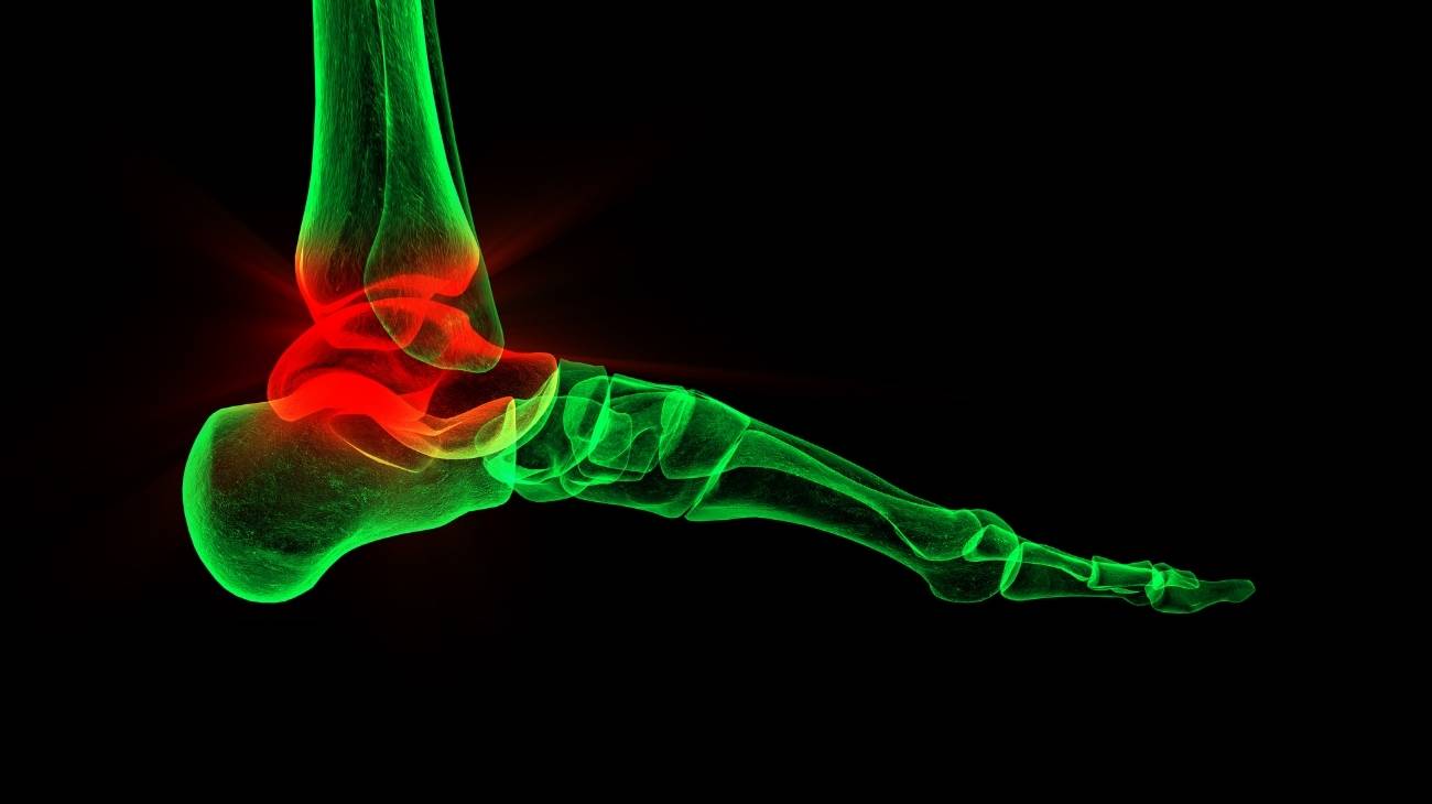
The ankle is an important joint for the traction of the foot on different surfaces and ground levels. Its anatomy makes it possible to coordinate the movements of the legs with those of the feet. This has a great effect on the occurrence of injuries, as it is an area that is very prone to inflammation and pain.
It is important to understand the anatomy of the ankle and the biomechanical movements it performs. This will help you to easily understand the ailments and diseases that are present. You can read this information below along with the treatments that combat ankle pain.
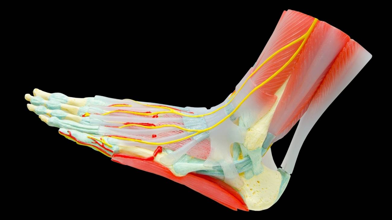
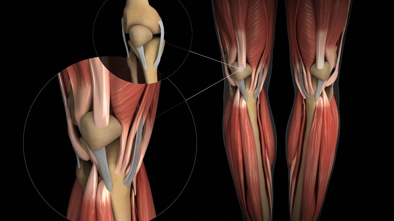
Parts and anatomy of the ankle
See below how the anatomy and different parts of the ankle are made up:
Bones and joints
The bones that make up the ankle joint are:
- Tibia: Connects at its internal malleolus with the talus and the external malleolus of the fibula. It works in coordination to perform movements in the foot, knee and hip.
- Fibula: This posterior leg bone connects at its external malleolus with the tibia, talus and calcaneus. Its job is to provide stability to the ankle.
- Astragalus: This is one of the bones that supports the weight of the body and transmits it to the foot, receiving it (in its upper part) from the malleoli of the tibia and fibula. In the lower lateral facet it is related to the calcaneus and the scaphoid. It is also called the talus.
- Calcaneus: This bone is what is known as the heel of the foot. It is responsible for articulating the cuboid, bearing the weight of the tibia and fibula, and stabilising the movements of the human body. It can also be considered part of the foot.
On the other hand, the joints that are present in the ankle are listed below:
- Astragalar trochlea or astragalar tibioperoneal: This is a joint that allows movement at the top of the ankle, between the talus of the foot and the tibia and fibula of the leg. Its cylindrical shape provides stability within the tibioperoneal mortise when flexion-extension movements of the foot are performed.
- Tibioperoneal mortise: It works in conjunction with the deltoid ligament whenever foot traction occurs and allows rotation of the fibula.
- Subtalar: This joint can also be considered part of the foot, as it is responsible for moving the talus and calcaneus. It is responsible for flexion and eversion movements of the foot.

Muscles
Learn about the muscle fibres that make up the ankle anatomy:
- Soleus: This broad muscle located at the back of the leg arises from the tibia and fibula and inserts into the calcaneus bone of the foot. It is directly related to the calf muscles and is responsible for joint flexion.
- Peroneus longus: It runs from the head of the fibula, at its external tuberosity, to the first metatarsal of the foot. Its action is the biomechanical movement of eversion.
- Peroneus brevis: Unlike the previous muscle, this tissue is located in the external part of the leg, in the lower part of the knee and is inserted in the fifth metatarsal. Its job is to perform plantar flexion, pronation and abduction of the foot.
- Flexor hallucis longus: It arises from the triceps suralis, the lower area of the fibula and extends to the distal phalanx of the big toe. Its name derives from the action it performs; that is, it flexes the hallux.
- Flexor digitorum longus: The function of this muscle is the same as that of the tissue mentioned for the big toe, but in this case it is responsible for flexing the other 4 tissues. It originates in the tibia and fibula and inserts in the proximal phalanges of each of the fingers.
- Gastrocnemius or calf muscle: Located at the back of the leg, above the soleus, it is responsible for flexing the sole of the foot. It starts at the femoral condyles, divides into three sections and inserts into the calcaneus.
- Anterior tibialis: It runs from the interosseous membrane, in the tibial area, to the second wedge (or medial cuneiform) and to the first metatarsal of the foot, in its second branch. Inversion and flexion of the foot is a consequence of this muscle.
- Posterior tibialis: Thanks to the work done by this muscle tissue, plantar flexion and adduction of the foot is possible. This is due to its elongated shape and posterior location in the leg. It originates in the proximal part of the tibia and on the medial side of the fibula, divides into three sections and ends in the dorsal area of the navicular, the medial cuneiform and the metatarsals.

Ligaments
The tendon bands located in the ankles are:
- Tibio-scaphoid: A tissue forming part of the deltoid ligament as a whole, which is incorporated within the collateral and tibial or medial ligaments. It arises from the tibial malleolus and inserts into the scaphoid.
- Tibiospring: It arises from the tibial malleolus in the anterior area of the tubercle and inserts into the plantar calcaneoscaphoid tissue, and is therefore considered a superficial ligament.
- Tibiocalcaneus: This is the last superficial ligament of the deltoid ligament complex. Like the other two, it arises from the tibial malleolus and inserts on the posterior part of the calcaneus.
- Tibiotalar: The only deep ligament of the deltoid connective tissue. It arises from the tibial malleolus and inserts on the medial tubercle of the talus.
- Anterior peroneoastotalar: This 3 by 20 millimetre ligament is part of the lateral peroneal connective tissue bundle. It arises from the lateral malleolus of the fibula and inserts on the talus.
- Posterior peroneoastotalar: It is also part of the lateral peroneal bundle and is the strongest. It is located in the lateral section of the ankle. It runs from the fibula at its distal end to the tubercle of the talus.
- Peroneocalcaneal: This is the last ligament that makes up the lateral peroneal ligamentous tissue group, but unlike the previous ones, it is not located inside the joint. It extends from the lateral malleolus of the fibula to the calcaneal tubercle.
- Intermalleolar: It can be divided into two sections, anterior and posterior. Its job is to allow dorsal flexion and plantar flexion of the foot by attaching the talus to the tibia. It is considered part of the distal tibioperoneal syndesmotic ligament complex.
- Transverse: It also belongs to the distal tibioperoneus. It runs from the malleolar fossa to the tibial malleolus. It provides the necessary balance in the movements of the foot.
- Anterior tibiofibular: Also known as the anteroinferior distal tibioperoneus, it is a ligament that joins the fibula to the tibia, leaving a space to produce articulation in the area.
- Tibioperonea distal posterior or tibiofibular posteroinferior: Like the other three ligaments, this connective tissue also belongs to the distal tibioperoneal syndesmotic ligament complex. It acts on the interosseous membrane of the tibia and fibula to provide stability of movement.
- Astragalus-calcaneus: It is responsible for generating the limits in the subtalar joint, since its route is carried out in the groove of the talus and calcaneus. It is part of the tarsal sinus ligaments and can be divided into medial and posterior.
- Cervical: This moment located between the talus and the medial surface of the calcaneus is responsible for balancing the inversion movements of the foot.

Best products for ankle and foot pain relief
Bestseller
-

2 Ankle Compression Sleeve (Black/Gray)
$19.95 -
2 Ankle Compression Sleeve (Green/Navy)
$19.95 -
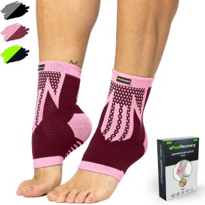
2 Ankle Compression Sleeve (Pink/Bordeaux)
$19.95 -
Electric Foot Massager
$58.97 -

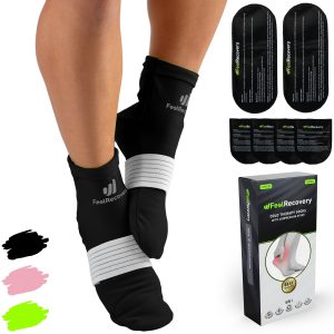
Ice Pack for Foot - Cold Therapy Socks (Black)
$24.95 -
Ice Pack for Foot - Cold Therapy Socks (Green)
$24.95 -
Ice Pack for Foot - Cold Therapy Socks (Pink)
$24.95 -
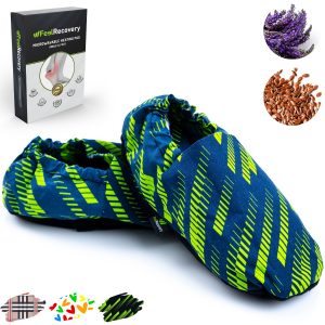
Microwavable Heated Slippers (Hearts)
$24.95 -
Microwavable Heated Slippers (Oxford)
$24.95 -
Microwavable Heated Slippers (Sport)
$24.95 -
Microwaveable Heating Pad for Pain Relief (Hearts)
$19.95 -
Microwaveable Heating Pad for Pain Relief (Oxford)
$19.95 -
Microwaveable Heating Pad for Pain Relief (Sport)
$19.95 -
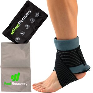
Reusable Gel Ice Packs for Ankle & Foot with Compression Band
$19.95 -
Reusable Gel Ice Packs with Compression Band for Sports Injury
$19.95 -
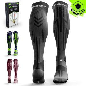
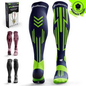
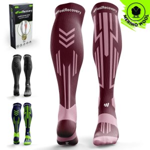
Sport Compression Socks (1 Pair) (Black/Gray)
$19.95 -
Sport Compression Socks (1 Pair) (Green/Navy)
$19.95 -
Sport Compression Socks (1 Pair) (Pink/Bordeaux)
$19.95


Biomechanics of the ankle
The different types of movements that the ankle can perform are related to the actions performed by the foot. Therefore, the biomechanics of the ankle comprise:
- Flexion: This movement consists of lifting the dorsal part of the foot in the direction of the tibia until the anterior edge of this bone touches the neck of the talus. The opening limit is 30°.
- Extension: This biomechanical action is the opposite movement of the section, causing plantar flexion.
- Rotation: This consists of rotating the tibia with the foot fixed, taking the malleoli as the axis. This action can be divided into internal and external, the former being the inward rotation of the body (or the other foot).
- Eversion: This action is the biomechanical movement in which the sole of the foot is raised towards the external side of the foot. Its amplitude is 25° with the body as the axis.
- Inversion: This is the internal movement opposite to eversion, but in this case the maximum opening limit is 35° from the axis.
Common ankle injuries
Types of ankle injuries
Among the most common ankle injuries suffered by a person are:
- Osteoarthritis of the ankle: This disease can be caused by different factors, the most common being trauma, demanding activities and overweight. It is a wear and tear that occurs in the articular cartilage, causing the bones of the ankle to collide with each other, resulting in pain and inflammation.
- Bursitis of the ankle: Bursae are sacs containing synovial fluid that keep the ankles cushioned when movements occur in this area. If there is an excess of this fluid, then the joint becomes inflamed causing swelling, paresthesia and lack of mobility.
- Ankle sprain: This is one of the most common diseases suffered by ligaments, especially when eversion and inversion movements occur. These are tears, microscopic or not, of the connective tissue. This causes bruising, pain and joint stiffness.
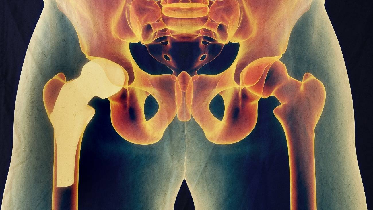
- Ankle fractures: Fractures that occur in the lower tibia and fibula and talus are not very common. They can be caused by blows or by bone diseases, for example, osteoporosis. Among these breaks, the most common are avulsion fractures.
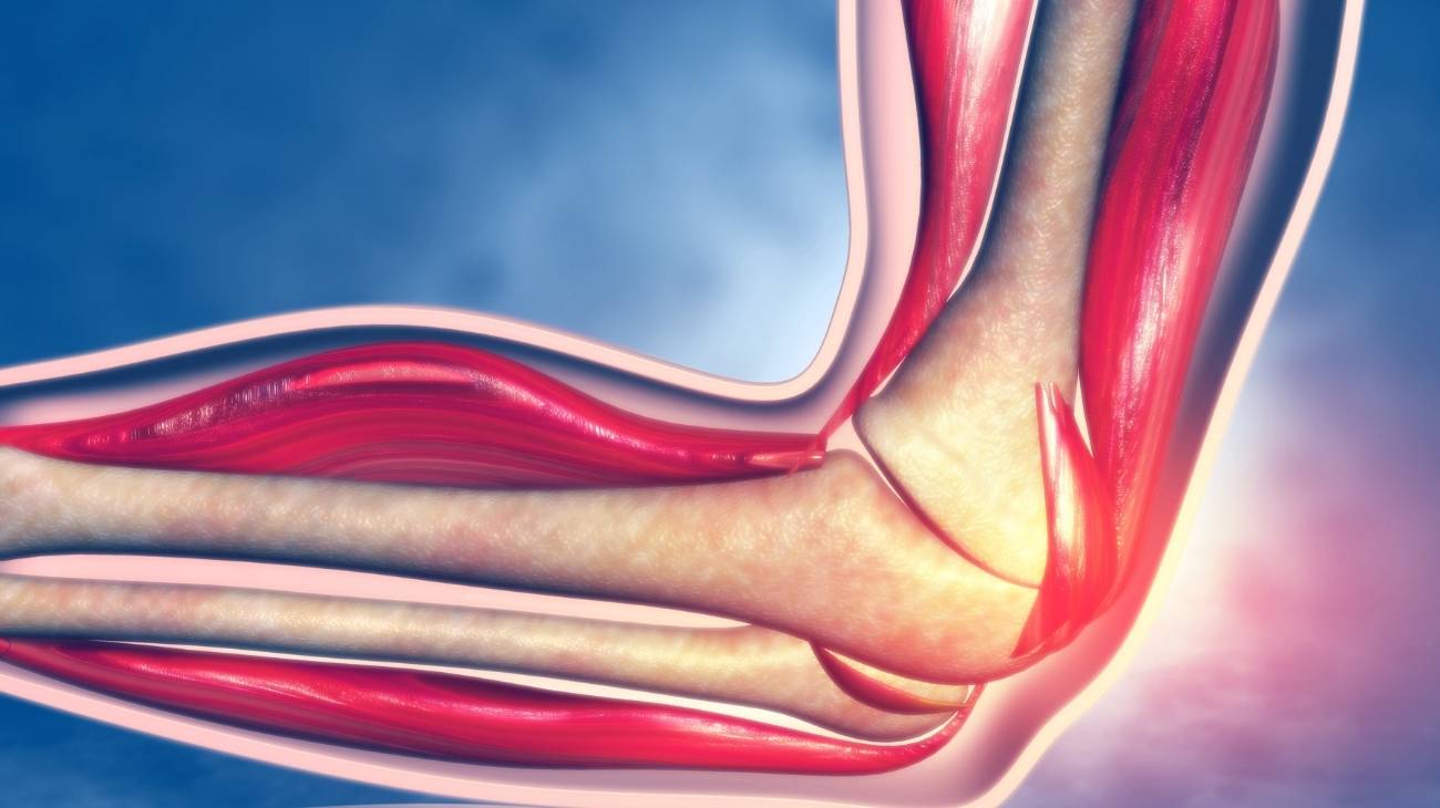
- Tendinitis of the ankle: Achilles tendinitis, posterior tibial tendinopathy and tarsal tunnel syndrome are the most common inflammations of the tendon tissues in the ankle. These conditions cause pain, joint stiffness, swelling and redness.
Sports injuries to the ankle
Sports are demanding activities that put the integrity of the ankle at risk. Find out which sports activities are most likely to injure this joint:
- Ankle injuries in badminton: Microscopic ligament tears and sprains are common, as are inflammations of the Achilles tendon due to repetitive movements. There are not many cases of contractures and ruptures.
- Foot, ankle and leg injuries in basketball: Syndesmosis sprains, soleus, peroneal and flexor muscle contractures are common. It is also possible to find athletes with tendinopathies.
- Ankle injuries in soccer: Fractures in the malleoli of the tibia and fibula are much more common than in other sports, although in reality sprains and achilles bursitis are the recurrent injuries in this sport.
- Foot and ankle injuries in gymnastics: Dislocations suffered by these athletes are common, but one cannot fail to mention inflammation of the bursae and wear and tear of the articular cartilage.
- Ankle injuries in running: The lateral and collateral ligaments are the most strained in this sport and are therefore prone to injury. Achilles tendinopathy, bursitis and muscle contractures can also be found.
- Ankle and foot injuries in tennis: Dislocations and sprains are common due to eversion and inversion movements of the foot. Some athletes may suffer from chronic Achilles tendonitis and osteoarthritis. Avulsion fractures may also occur.
- Volleyball ankle injuries: Sprains up to fourth degree are recurrent in volleyball. The Achilles tendon is also easily inflamed, but dislocations and contractures in the soleus and calf muscles are the most commonly seen injuries.
- Ankle injuries in Yoga: Achilles tendonitis and tears are the most common injuries that occur in this activity, in a few cases people are found with inflamed bursae and joint immobility due to inflammation of ligaments.
Diseases and ailments of the ankles

Among the diseases and ailments that frequently appear in the ankle, we can mention the following:
Osteoporosis
The density of the bone mass is affected in the bones of the ankle. This causes a decrease in the stiffness of these tissues (generated by the malfunctioning of the cells in the central part of the bones). A person suffering from this condition is prone to ankle fractures and osteophytes, which can lead to osteoarthritis and other ailments.
Varicose veins
This disease causes varicose veins to become enlarged, which causes problems in the transfer of oxygen-depleted blood to the body. This causes leg pain and tiredness, especially in the ankle area.
Gout
This condition is caused by excess uric acid in the ankle that cannot be naturally secreted by the blood. This condition causes microcrystals to form in this part of the body, which can lead to overbones and osteophytes. This is considered a risk factor for osteoarthritis.
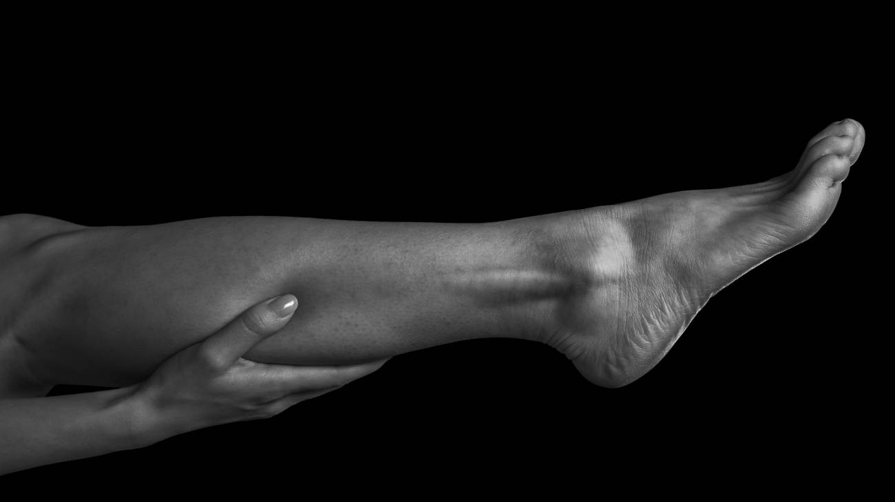
How can we relieve ankle and leg pain through complementary and non-invasive therapies?
To alleviate pain and other symptoms in the ankles, it is advisable to resort to these complementary treatments:
Heat and cold therapy
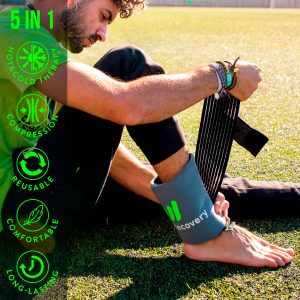
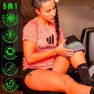
It is possible to reduce inflammation and pain in the ankles by means of this non-invasive complementary therapy. It involves applying different products that allow the benefits of cold and heat to be obtained. To reduce swelling and improve blood flow , it is advisable to use cold gel packs with compression straps to maintain the ideal temperature in the affected area. Consult your doctor to choose the best product.
Compression therapy
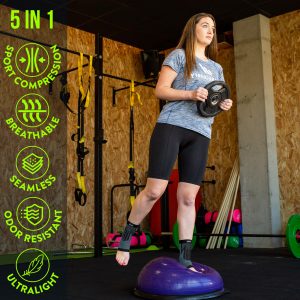
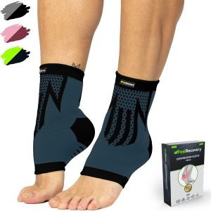
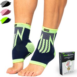
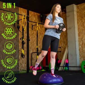
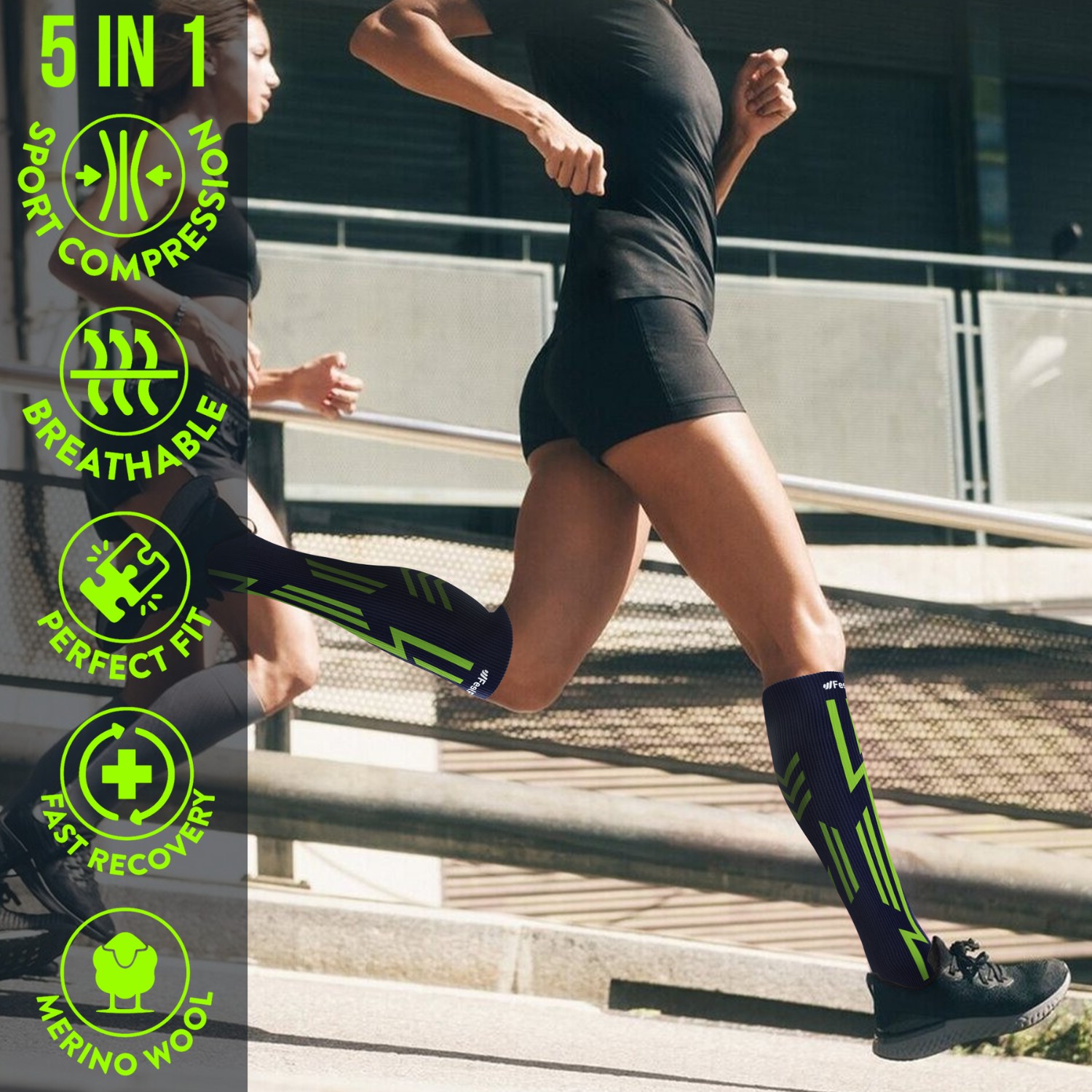
Symptoms of ankle pain and swelling may improve and go into remission if compression therapy is applied with compression garments and products. These products will help to improve the exchange of nutrients from the blood to remove any remaining metabolites that may be in the joint. There are different techniques for this non-invasive therapy, but the most effective is the use of compression ankle braces, sports calf sleeves and compression socks. You can choose the most appropriate product based on your doctor's advice.
Massage therapy
One of the most commonly used therapies for foot and ankle complaints is massage therapy. This technique consists of improving the dilation of the capillary vessels so that blood flows in the affected area. You can choose a professional masseur, but if you want to save money you can find products that will help you with this task. For the latter option you will find massage rollers, massage guns and electric foot massagers, among others.
Acupressure therapy
This non-invasive technique has been applied for hundreds of years in China, so its results are guaranteed to improve blood flow in the ankles and thus reduce pain and inflammation. It consists of massaging by means of constant controlled pressure on different parts of the body, using massage rollers, massage balls and acupressure pads.
Thermotherapy
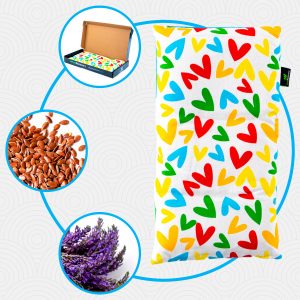
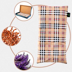
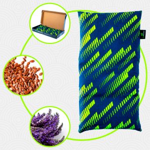
One of the most commonly used non-invasive therapies to reduce pain and stiffness in the ankles is the application of heat directly to this part of the body. Different techniques can be used for this purpose, the most practical and efficient being the use of microwavable thermal pillows or packs with heat gels.
Cryotherapy
To improve inflammations and to achieve a quick mobility of the ankle, the application of thermal pillows and cold gels packs is convenient. These products can be used directly on the joint to achieve anti-inflammatory and sedative effects. Sessions should not exceed 15 to 20 minutes and should be done three to four times a day.
Electrical muscle stimulation (EMS)
Muscle electrostimulation, or EMS, is a therapy that consists of stimulating muscle contractions through the use of electricity, so as to achieve an effect of activity and hypertrophy as in the gym, but without the need to go to any sports center. This means that you can put your muscles to work without leaving home.
Electrotherapy
This is a technique that seeks relief from pain and some physical ailments through the application of electrical and electromagnetic energy, among other variants, through the skin with the use of conductive pads called electrodes. It is a very safe type of therapy and must be applied by a physical therapist specialized in the manipulation of electricity to treat some kinds of ailments.
Myofascial release therapy
Also known as myofascial induction, this therapy consists of the application of manual massage to treat the shortening and tension generated in the myofascial tissue that connects the muscles to the bones and nerves. For this purpose, various massage techniques are used that focus on the so-called trigger points.
Percussion Massage Therapy
Vibration or percussion massages are precise, rhythmic and energetic strokes on the body to achieve relief from some annoying symptoms when muscle fibers are tightened, often by a high workload on them and that has left trigger points in the muscle fibers.
R.I.C.E Therapy
The R.I.C.E. therapy is the first and simplest of the treatment protocols for minor injuries. It appears in the sports field to deal with accidents involving acute injuries. For many years, it has been considered the most suitable for its speed and results.
Trigger points therapy
Myofascial pain points or trigger points are knots that are created in the deeper muscle tissues, causing intense pain. The pain does not always manifest itself right in the area where the point develops, but rather this pain is referred to nearby areas that seemingly do not appear to be related. In fact, it is estimated that more than 80% of the pain they cause manifests in other parts of the body.
Other effective alternative therapies
It is possible to apply not only the therapies mentioned above to relieve pain, but there are also other non-invasive treatments that are effective in healing ankles.
- Natural remedies with the use of plants: One of the least invasive therapies within the complementary treatments is the use of plants with medicinal effects. It is possible to add sage, rosemary and sage to a bath with warm water to obtain anti-inflammatory benefits.
- Acupuncture: Through this therapy the patient finds mental harmony thanks to the nerve stimulation caused by needles placed in different areas of the body.
- Kinesiotherapy: Through short, repetitive movements, the ankle joint is stimulated to recover its biomechanical functions. It is carried out under strict professional control.
- Aromatherapy: The application of different aromas of citrus fruits or certain plants causes the patient to relax and reduces stress. This helps to cope better with ankle pain.
References
- Kelikian, A. S., & Sarrafian, S. K. (Eds.). (2011). Sarrafian's anatomy of the foot and ankle: descriptive, topographic, functional. Lippincott Williams & Wilkins. https://books.google.es/books?hl=en&lr=&id=I8h6bDR0SLMC
- Golanó, P., Vega, J., De Leeuw, P. A., Malagelada, F., Manzanares, M. C., Götzens, V., & Van Dijk, C. N. (2010). Anatomy of the ankle ligaments: a pictorial essay. Knee Surgery, Sports Traumatology, Arthroscopy, 18(5), 557-569. https://link.springer.com/article/10.1007/s00167-010-1100-x
- Brockett, C. L., & Chapman, G. J. (2016). Biomechanics of the ankle. Orthopaedics and trauma, 30(3), 232-238. https://www.sciencedirect.com/science/article/pii/S1877132716300483
- Riegger, C. L. (1988). Anatomy of the ankle and foot. Physical therapy, 68(12), 1802-1814. https://academic.oup.com/ptj/article-abstract/68/12/1802/2728302
- Tropp, H. (2002). Commentary: functional ankle instability revisited. Journal of athletic training, 37(4), 512. https://www.ncbi.nlm.nih.gov/pmc/articles/PMC164386/
- Garrick, J. G., & Requa, R. K. (1988). The epidemiology of foot and ankle injuries in sports. Clinics in sports medicine, 7(1), 29-36. https://www.sciencedirect.com/science/article/abs/pii/S027859192030956X
- Glick, J. M., Gordon, R. B., & Nishimoto, D. (1976). The prevention and treatment of ankle injuries. The American journal of sports medicine, 4(4), 136-141. https://journals.sagepub.com/doi/abs/10.1177/036354657600400402?journalCode=ajsb
- Fong, D. T. P., Hong, Y., Chan, L. K., Yung, P. S. H., & Chan, K. M. (2007). A systematic review on ankle injury and ankle sprain in sports. Sports medicine, 37(1), 73-94. https://link.springer.com/article/10.2165/00007256-200737010-00006
- Giza, E., Fuller, C., Junge, A., & Dvorak, J. (2003). Mechanisms of foot and ankle injuries in soccer. The American journal of sports medicine, 31(4), 550-554. https://journals.sagepub.com/doi/abs/10.1177/03635465030310041201
- Polzer, H., Kanz, K. G., Prall, W. C., Haasters, F., Ockert, B., Mutschler, W., & Grote, S. (2012). Diagnosis and treatment of acute ankle injuries: development of an evidence-based algorithm. Orthopedic reviews, 4(1). https://www.ncbi.nlm.nih.gov/pmc/articles/PMC3348693/