The skull and face are responsible for protecting the brain, a fundamental organ for human development. Headaches are common in a large number of people, while the causes and symptoms that produce them can vary depending on which part of the body is not working properly.
For this reason, it is important to know the anatomy of the head, eyes and face and the most common diseases that occur in this part of the body. You will find this information below, so we invite you to read on and have a look!
Parts and anatomy of the head
The anatomy of the head is made up of the following:
Skull
The skull is made up of the following bones:
- Frontal: The bone in the anterior and superior view of the skull. It is a central, symmetrical bone that is joined to the parietal, sphenoid, nasal, ethmoid, zygomatic, maxillary and lachrymal bones.
- Parietal: Two bones located in the anterolateral part of the skull. It is a large bone that covers a large part of the brain. It connects with the frontal, sphenoid, temporal and occipital bones,
- Temporal: Like the parietal bone, it is an even bone, but it is located in the anterior part of the skull. It is connected to the frontal, temporal, parietal and zygomatic bones of the face. It is located at the level of the temple.
- Occipital: This bone forms the base part of the skull, which connects to the atlas or first cervical vertebra of the spine. It also connects with the parietal and temporal bones.
- Sphenoid: This is a deep, odd bone located at the middle base of the skull. It connects with the frontal, temporal and occipital bones. The pituitary glands and meninges lie within this endocranial recess.
- Ethmoid: Its T-shape allows connection with the frontal, sphenoid, maxillary, nasal, lacrimal and facial mass.
The muscles that make up the skull are
- Occipitofrontal: This muscle is located in the posterior area of the head, from the epicranial fibrous membrane to the mastoid process of the temporalis. It is also known as the occipital belly.
- Temperoparietal: The temporalis fascia is covered by this muscle, which arises from the upper part of the skull (precisely in the aponeurotic galea) to its insertion in the helix of the ear.
- Sternocleidomastoid: This is a flat muscle located at the base of the temporal bone and develops to the sternal manubrium.
- Procerus: This muscle is responsible for moving the eyebrows downwards. It arises from the lower part of the frontal bone and inserts into the eyebrows.
- Superciliary corrugator: It is possible to wrinkle the forehead thanks to the action of this muscle. Its trajectory runs from the superciliary arch to the eyelid skin.
- Temporalis: Also known as crotaphites, it is a muscle that allows the elevation and retraction of the jaw bone. It runs from the inferior temporalis to the process of the lower jaw.
The joints that exist in the skull are of the synarthrosis type. That is to say, they have a small membrane or no movement between the bones.

Face
The bones of the face are
- Upper jaw: It occupies a large portion of the face, is an even bone and is quadrilateral in shape. It is found in the central anterior part of the bones of the face, connecting with the zygomatic, frontal, lacrimal, nasal, ethmoid, vomer palatine and inferior turbinate. It gives rise to the upper teeth and molars.
- Lower jaw: It is also known as the mandible. It is the largest bone of the face, it is a pair and symmetrical. It has the joint with the most movements in the face in its posterior part. It is responsible for holding the teeth and molars underneath.
- Nasal: This paired bone is joined together by the septal cartilage, forming the external part of the nose. Its upper side connects with the frontal bone and the inner side with the ethmoid bone.
- Vomer: Part of the nasal septum, it is a single bone that divides the nose into two nostrils. It joins the upper jaw, the palatines, the ethmoid and the sphenoid.
- Turbinates: They can be divided into upper, middle and lower turbinates and form a bony structure inside the nose. They are located on the lateral side of the nostrils.
- Zygomatic or Malar: Constitutes part of the ocular orbit and the cheekbone of the face. It is a protruding bone that joins the maxilla, sphenoid, frontal and temporal bones.
The following muscles are found in the face:
- Orbicularis oculi: This can be divided into the orbital portion and the palpebral portion. This muscle arises from the frontalis, passing through the lacrimal canal to the skin of the eyelids. Its function is to close the eyelids.
- Nasal: This muscle can be found on the lateral aspect of the nose, as it originates in the upper jaw and inserts into the nasal bone. It elevates the turbinate bones of the nostrils and controls the movements of the tip of the nose.
- Levator labii superioris: This muscle is named after the action it performs. It arises from the infraorbital rim and inserts into the skin and muscle located in the upper lip.
- Lesser zygomatic muscle: This muscle of the face is a small tissue that arises from and inserts into the external face of the malar or zygomatic bone.
- Greater zygomatic: It runs from the temporal process to the corner of the mouth. Its job is to pull the skin around the mouth.
- Buccinator: This is a flat muscle located on the cheek of the face, it acts in mastication and compresses the cheeks. It originates in the upper jaw and inserts into the orbicularis oris muscle.
- Orbicularis oris of the mouth: The lips can be folded thanks to the action of the muscle, which has a trajectory from the upper jaw to the skin located in the labial commissure.
- Masseter: Its action allows the mouth to be closed and jaw movements to be controlled. This muscle runs from the zygomatic process to the anthropometric point of the mandible.
- Risorius: A small muscle located next to the orbicularis oris muscle of the mouth, it is characterised by retracting the corner of the mouth, which is part of the action of laughing in the person.
- Depressor labii inferioris: Like other muscles, this tissue is named after the action it performs. It develops from the lower jaw to the skin of the lower lip.
- Mentonian: It is responsible for lifting the skin on the chin. It arises and inserts in the lower part of the lips.
- Mouth angle depressor: The corner of the mouth can be depressed thanks to the action of this muscular tissue. It arises from the chin and inserts into the corner of the mouth.
- Platisma: It is responsible for lowering the jaw and tightening the skin when certain muscles of the face contract. It is a large muscle that originates in the subcutaneous part of the clavicle and inserts in the chin region.
- Pterygoid: A group of muscles responsible for chewing that arises from the sphenoid to the mandible, causing its depression and elevation.
Ligaments that can be found in the face are:
- Sphenomandibular: This ligament allows the sphenoid to join the mandibular foramen and the maxillary vessels.
- Lateral ligament: This ligament is divided into two sections that allow the posterior aspect of the neck of the maxillary bone to be joined to the zygomatic bone.
- Pterygospinous: The spinous process of the sphenoid bone is joined to the lateral pterygoid by this tissue.
- Stylomandibular: Another short, thin ligament that allows the sphenoid to attach to the mandible.
- Stylohyoid: This is one of the longest ligaments that can be found on the face, as it facilitates the attachment of the hyoid bone to the styloid process of the temporal bone.
The main joints found in the face are
- Temporomandibular: This articular body is responsible for carrying out the movements between the mandible and the temporal bone. It is of the synovial type.
- Synarthrosis type joints.

Eyes
The orbital cavity is anatomically formed by these bones:
- Lacrimal, lacrimal or unguis: This is a square-shaped plate that articulates on each of its four sides with the frontal, ethmoid, nasal and maxillary bones.
- Ethmoid: As mentioned above, this T-shaped bone creates the side of the orbital cavity of the eye.
- Other bones already discussed in the face: zygomatic, maxillary, palatine and sphenoid.
The muscles of the eyes are:
- Greater oblique: Allows movement of the eye in all directions, its course is from the back of the eye to the cartilaginous ring of the orbit.
- Lesser Oblique: Also part of the extrinsic musculature of the eye, but unlike the other muscles it inserts into the common tendinous ring or "Ring of Zinn".
- Superior rectus: Its name refers to the location of this muscle, at the top of the ocular orbit. It enables eye rotation and elevation movements.
- Rectus internus: This muscle tissue allows contraction of the eye socket, allowing the eye to turn in the direction of the nose.
- External rectus: The action of this muscle allows the eyeball to rotate in the opposite direction to the nose, known as abduction of the eye.
- Inferior rectus: This is the last of the group of six muscles that balance the movements of the globe. This tissue is responsible for rotating the eye downwards and allowing movement towards the middle of the eye.
The ligaments found in the eye are:
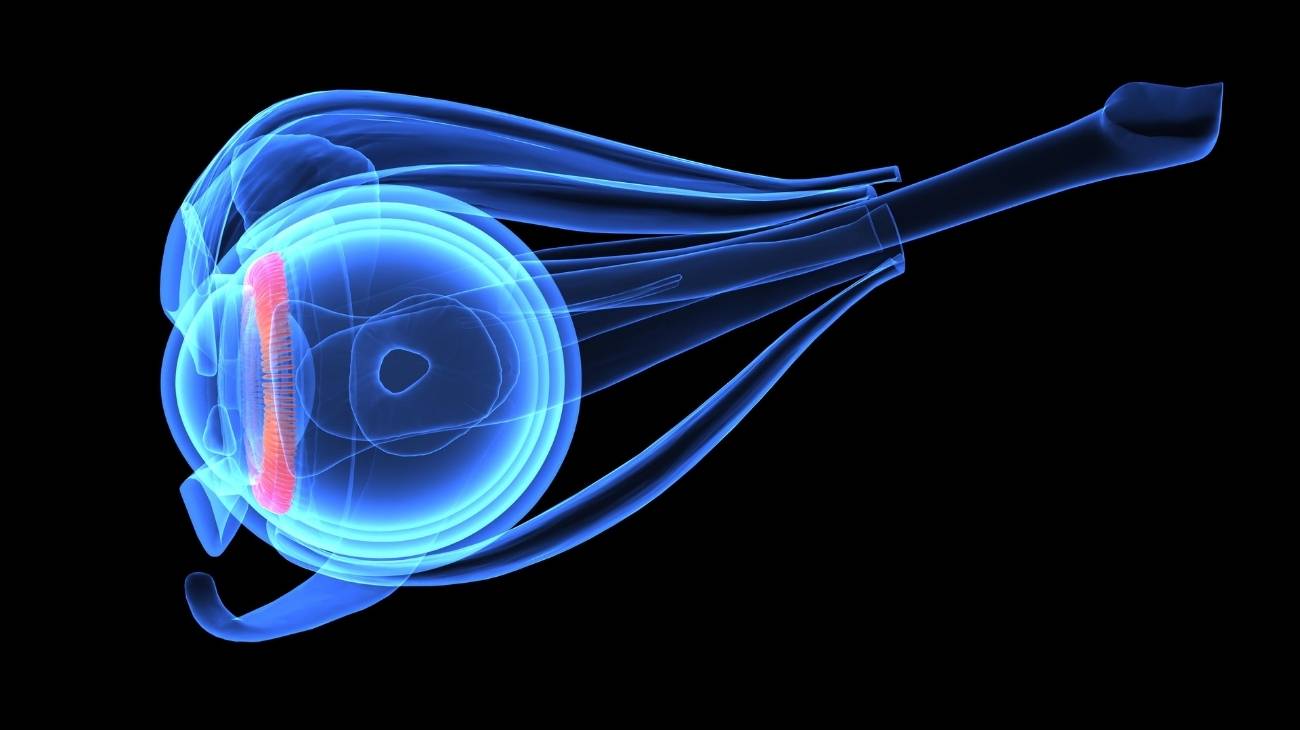
- Lens suspensory: Also known as the ciliary zonule. This ligament made of carbohydrates and proteins stabilises the lens of the eye to improve focusing on near objects.

Best products for head, face and eye pain relief
Bestseller
-

Acupressure Mat and Pillow (Black/Gray)
$49.95 -
Acupressure Mat and Pillow (Green/Navy)
$49.95 -
Acupressure Mat and Pillow (Pink/Bordeaux)
$49.95 -
Acupressure Pillow (Black/Gray)
$29.46 -
Acupressure Pillow (Green/Navy)
$29.46 -
Acupressure Pillow (Pink/Bordeaux)
$29.46 -
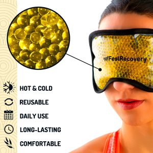
Gel Eye Mask for Puffy Eyes (Gold/Black)
$11.95 -

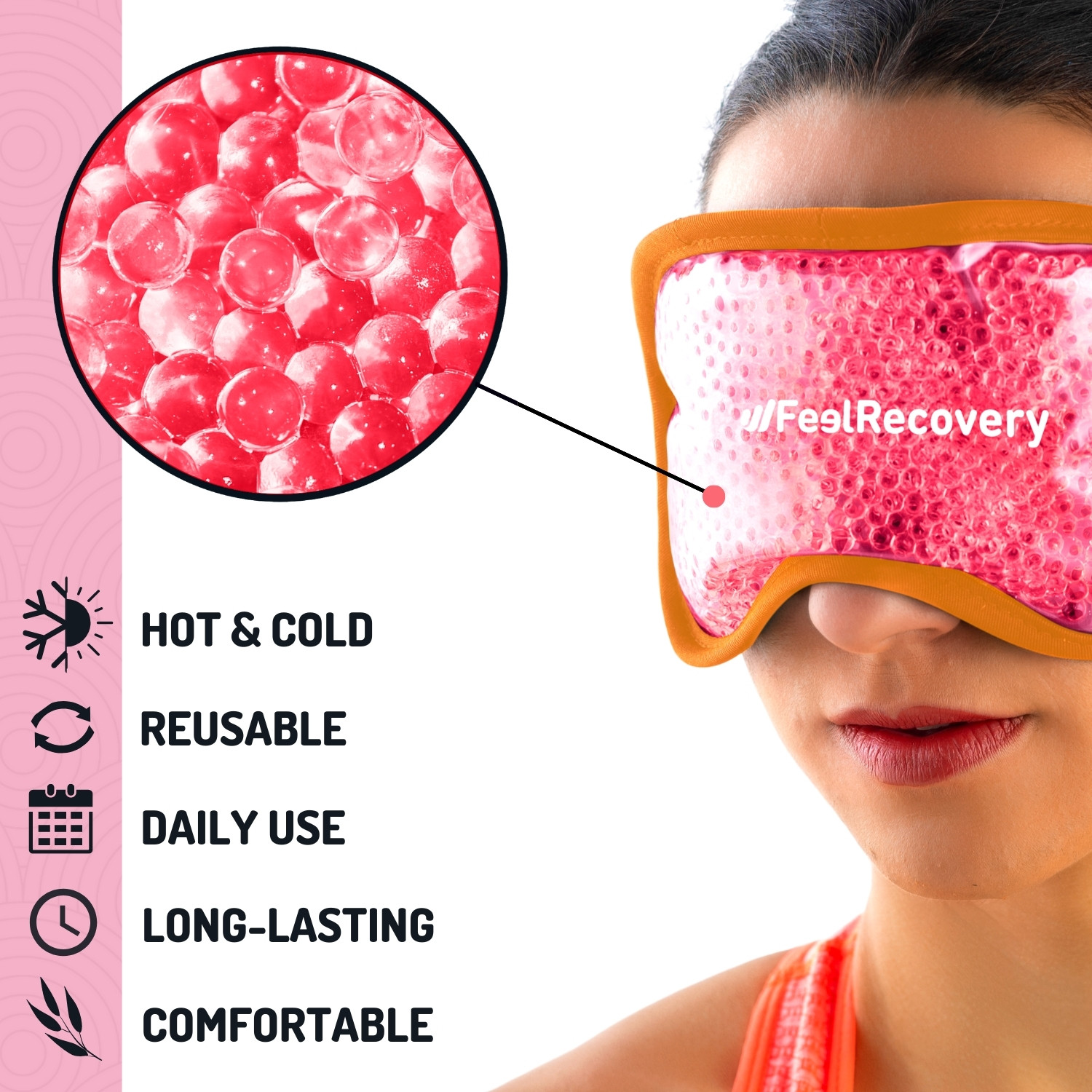
Gel Eye Mask for Puffy Eyes (Orange/Pink)
$11.95 -
Gel Eye Mask for Puffy Eyes (Purple/Turquoise)
$11.95


Most common eye diseases and ailments
The following are the most common eye diseases and ailments:
Myopia
This disease results in a person's inability to see distant objects clearly, which causes blurred vision. There are different causes that provoke this eye condition, but the main one is hereditary.
Astigmatism
The irregular curvature of the crystalline lens of the eye causes blurred vision of near and distant objects (in some cases there is a perception of distorted images). It can occur at different ages, as this disease is usually hereditary.
Presbyopia
Presbyopia is one of the most common eye diseases in men between 40 and 45 years of age, and can increase up to the age of 65. It is the inability to focus on close objects, with one of its characteristic symptoms being the need to read at a certain distance, usually with the arm fully extended.
Conjunctivitis
This contagious disease causes itching, eye discharge and redness in the eye due to the presence of viruses or bacteria that inflame the conjunctiva (small layer that covers the white part) of the globule.
Cataracts
In addition to producing blurred vision due to the opacity of the crystalline lens of the eye, people can also perceive objects in a double way. There are different causes of cataracts, and not the most common is the advanced age of the patient.
Glaucoma
There are different types of glaucoma, the most common being open-angle glaucoma, which consists of a gradual loss of sight. It is a disease caused by high pressure in the eye, which can appear at any age, although it is common to find patients over 60 years of age.
How can we relieve headache pain through complementary and non-invasive therapies?

See below the complementary treatments that are applied to relieve head, face and eye pain:
Heat and cold therapy
This type of treatment consists of applying heat in the same session, then continuing with cold and finishing with the first temperature. Care must be taken, as the application should not exceed 20 minutes in total.
- Heat causes the vessels in the capillary walls to dilate, resulting in increased blood flow to the affected area, and the use of microwavable heat packs is recommended.
- While cold acts as an analgesic agent. It is possible to apply this therapy by means of ice gel packs.
Massage therapy
Massages stimulate the blood vessels to improve blood circulation in the affected area. In addition, it reduces stress because the body itself generates dopamine causing a feeling of well-being. This can help to reduce headaches. Electronic massagers, vibration rollers, massage guns and electric back massage chairs can be used to help reduce headaches.
Acupressure therapy
Compression is a massage technique that is applied to specific places on the body to reduce headaches. For this reason, neck, back and shoulder massage cushions or vibration balls can be used in this therapy. This will stimulate the capillary walls to improve blood flow.
Thermotherapy
Although we have already talked about the benefits of heat as a dilating agent of the capillary walls, it is important to clarify at this point that this therapy uses only heat to obtain these effects (unlike heat and cold treatment). Thermotherapy can be applied by means of different techniques, the most common being the use of microwavable thermal pillows and hot water baths.
Cryotherapy
Care must be taken with the application of cold, either by means of cold pads or ice gel packs, because the effect that cryotherapy can generate may be contraindicated for headaches. In other words, the use of this therapy must be recommended by the doctor and is only possible when the injury has not exceeded 48-72 hours.
Electrotherapy
This is a technique that seeks relief from pain and some physical ailments through the application of electrical and electromagnetic energy, among other variants, through the skin with the use of conductive pads called electrodes. It is a very safe type of therapy and must be applied by a physical therapist specialized in the manipulation of electricity to treat some kinds of ailments.
R.I.C.E Therapy
The R.I.C.E. therapy is the first and simplest of the treatment protocols for minor injuries. It appears in the sports field to deal with accidents involving acute injuries. For many years, it has been considered the most suitable for its speed and results.
Other effective alternative therapies
There are other therapies that can be applied due to their non-invasive methods. These are:
- Natural remedies with the use of plants: Through this treatment it is possible to stimulate different areas of the body that will cause the headache to diminish, if it is caused by a body injury or by a hepatic or gastric pathology. Boldo, chamomile and linden are often used.
- Acupuncture: This oriental technique allows the patient to relax, seeking mental balance. If the headache is caused by a contusion or extreme pain in any part of the body, with this therapy it is possible to reduce these symptoms.
- Aromatherapy: This is a therapy that consists of relaxing the patient by means of aromas that reduce stress, thus improving the headache. It can be applied by means of essential oils or air diffusers. Among the most common herbs are lavender, peppermint and peperine.
References
- Wang, Y. X., Panda-Jonas, S., & Jonas, J. B. (2021). Optic nerve head anatomy in myopia and glaucoma, including parapapillary zones alpha, beta, gamma and delta: histology and clinical features. Progress in retinal and eye research, 83, 100933. https://www.sciencedirect.com/science/article/pii/S1350946220301051
- Mackenzie, P. J., & Cioffi, G. A. (2008). Vascular anatomy of the optic nerve head. Canadian Journal of Ophthalmology, 43(3), 308-312. https://www.sciencedirect.com/science/article/abs/pii/S0008418208801111
- Willoughby, C. E., Ponzin, D., Ferrari, S., Lobo, A., Landau, K., & Omidi, Y. (2010). Anatomy and physiology of the human eye: effects of mucopolysaccharidoses disease on structure and function–a review. Clinical & Experimental Ophthalmology, 38, 2-11. https://onlinelibrary.wiley.com/doi/full/10.1111/j.1442-9071.2010.02363.x
- Radius, R. L., & Gonzales, M. (1981). Anatomy of the lamina cribrosa in human eyes. Archives of ophthalmology, 99(12), 2159-2162. https://jamanetwork.com/journals/jamaophthalmology/article-abstract/634026
- Kels, B. D., Grzybowski, A., & Grant-Kels, J. M. (2015). Human ocular anatomy. Clinics in dermatology, 33(2), 140-146. https://www.sciencedirect.com/science/article/abs/pii/S0738081X1400234X
- Carpenter, W. B. (1866). Principles of human physiology. Lea & Blanchard. https://books.google.es/books?hl=en&lr=&id=6L40AQAAMAAJ
- Bentsianov, B., & Blitzer, A. (2004). Facial anatomy. Clinics in dermatology, 22(1), 3-13. https://www.sciencedirect.com/science/article/abs/pii/S0738081X03001135
- Marur, T., Tuna, Y., & Demirci, S. (2014). Facial anatomy. Clinics in dermatology, 32(1), 14-23. https://www.sciencedirect.com/science/article/abs/pii/S0738081X13000898