The hip is one of the most important joints for supporting the body's weight during biomechanical movements and when standing. This is due to the anatomy of this joint area, in which a series of ligaments and muscles come together to act precisely with the bones.
From this, it is known that the hip is highly susceptible to injury. For this reason, it is important that you know which are the most frequent ailments of this joint and how to treat them. Take a look at all the information we have prepared for you in a simple and precise way.
Parts and anatomy of the hip
Below, we will show you the parts and human anatomy of the hip:
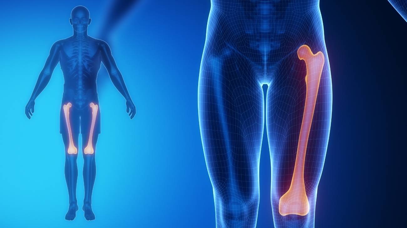
Bones and joints
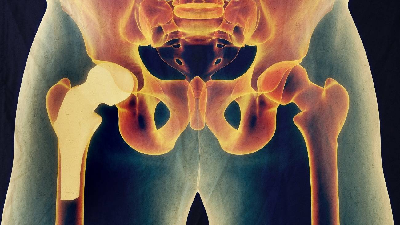
The bones that make up the hip are the ilium (the upper part of which is known as the iliac crest), the sciatic spine, the ischium, the sacrum, the pubis, the acetabulum and the coccyx. The sciatic spine, in women, is displaced in relation to the pelvic cavity, the latter being much more open in the case of women so as not to obstruct the birth canal.
Within the hip are also the sacroiliac and coxofemoral joints. The first joint connects the iliac crest to the lumbar spine, forming the characteristic movements of the lower back. The second joint connects the head of the femur to the socket of the acetabulum, enabling the person to walk and perform other movements according to the amplitude of this joint body.

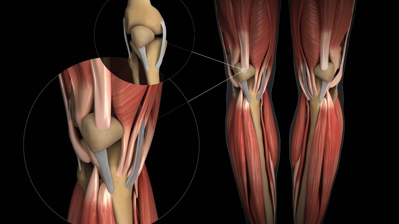
Muscles
The muscles that are part of the hip anatomy are the following
- Piriformis: The purpose of this muscle is to allow external rotation, extension and adduction at the hip. It originates from the sacrum and inserts into the greater trochanter at the upper edge.
- Psoas iliacus: Located in the anterior area of the thigh, inside the abdominal cavity. The work of this muscle is to move the trunk and produce hip flexion. The internal iliac fossa and the lumbar vertebrae are its origin, while the trochanter of the femur is its insertion site.
- Tensor of the fascia lata: It is a thin, flattened muscle found on the gluteus. Its function is to stabilise hip movements and knee extension. It arises from the iliac spine and inserts into the lateral tendons of the fascia lata called the iliotibial band.
- Adductor: This muscle consists of three parts named adductor minimus, medius and adductor magnus. It originates from the pubis and ischium and inserts into the femur. Its job is to flex, rotate and stabilise the spine and pelvis.
- The gracilis: It is also known as the internal rectus and has several insertions, which are the symphysis pubis, the medial condyle of the tibia and some internal tendons. Internal rotation, abduction and flexion of the hip are the functions of this muscle.
- Pectineus: The activity of this muscle is to move the leg together with the adductors. It inserts on the femur in the area of the pectineus line and originates from the iliopubic ramus.
- Rectus femoris: It arises from the iliac spine and inserts on the quadriceps tendon. Knee extension and hip flexion are the two actions performed by this muscle.
- Gluteus: Like the adductor, it can be divided into three sections, named gluteus maximus, gluteus medius and gluteus minimus. They are located in the anterior part of the hip and insert on the edge of the iliotibial tract muscles and on the gluteal tuberosity. They vary in strength being the sides and edges of the sacrum and coccyx, as well as the sacrotuberous ligaments.
- Geminus superior and geminus inferior: They are also known as the superior gemellus and inferior gemellus. They are found in the gluteal and femoral regions of the body whose job is to adduct the hip and cause movements in the coxofemoral joint body.
- Quadratus femoris: Also known as the quadratus cruris, it travels from the ischium to the intertrochanteric crest. Its action is to rotate the femur laterally.
- Obturator internus: Named after its origin in the obturator foramen of the hip, it inserts into the trochanter of the femur and into the femoral fossa. Its action allows external rotation of the hip.
- Obturator externus: The external margin of the obturator foramen is the origin of this muscle, and its insertion is into the tendon of the femoral neck. Abduction of the hip is caused by this muscle.
- Sartorius: It is the longest muscle in the human anatomy and its course is on the superficial part of the thigh. Its job is to flex the hip and move the femur away from the femur via the adductors, which creates the ability to walk or place a heel on a line above the opposite knee.

Ligaments
Within the structure of the hip it is possible to find an arrangement of ligaments that help the functioning of this part of the body.
These tissues are:
- Round or femoral head ligament: It runs from the acetabulum to the fossa in the femoral head. This allows the coxofemoral joint to function.
- Iliofemoral: This ligament divides in its lower part in two, causing an inverted "Y" shape. It arises from the iliac spine and inserts into the anterior area of the femur, within the trochanter line (in two different areas, hence the name for each subdivision, Superior and Inferior).
- Pubofemoral: Its origin is at the top of the pubis and inserts into the lower section of the ischiofemoral, taking into account the intertrochanteric line.
- Ischiofemoral: its origin is the ischium, running along the posterior part of the acetabulum bone until it ends a little below the head of the femur.
- Sacrotuberous: It is considered as a continuation of the biceps femoris, its origin is in the upper area of the coccyx, in the tubercles of the sacrum and in the lower area of the sacrum. It joins the iliac spine with other ligaments.
- Sacrospinous: This ligament runs from the ischial spine to the edges of the coccyx and sacrum. Its function is to produce hip movements and to strengthen other ligaments.

Best products for hip and lower back pain relief
Bestseller
-

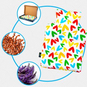
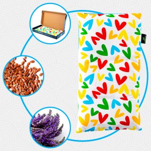
Heating Pad for Microwave Classic Bottle Shaped (Hearts)
$24.95 -
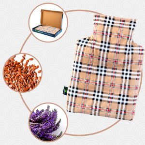
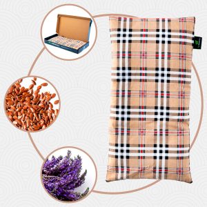
Heating Pad for Microwave Classic Bottle Shaped (Oxford)
$24.95 -
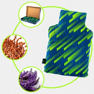
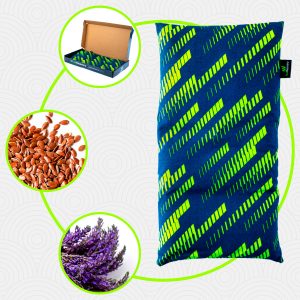
Heating Pad for Microwave Classic Bottle Shaped (Sport)
$24.95 -

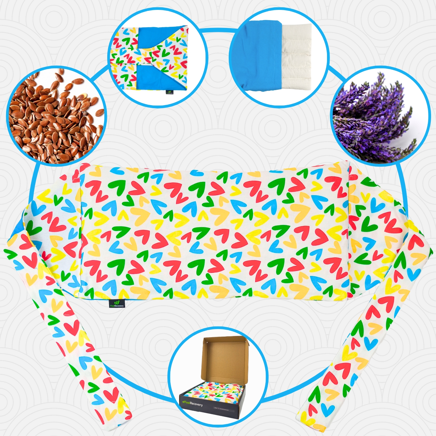
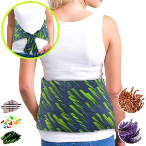
Microwave Heating Pad for Back Pain Relief (Extra Large) (Hearts)
$29.95 -
Microwave Heating Pad for Back Pain Relief (Extra Large) (Oxford)
$29.95 -
Microwave Heating Pad for Back Pain Relief (Extra Large) (Sport)
$29.95 -
Microwave Heating Pad for Neck & Shoulder Pain Relief (Hearts)
$29.95 -
Microwave Heating Pad for Neck & Shoulder Pain Relief (Oxford)
$29.95 -
Microwave Heating Pad for Neck & Shoulder Pain Relief (Sport)
$29.95 -
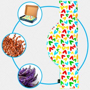
Microwaveable Heating Pad for Pain Relief (Hearts)
$24.95 -
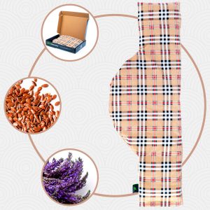
Microwaveable Heating Pad for Pain Relief (Oxford)
$24.95 -
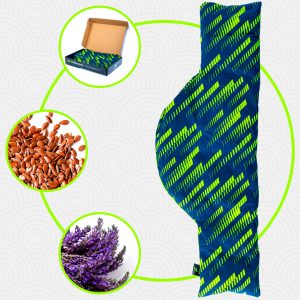
Microwaveable Heating Pad for Pain Relief (Sport)
$24.95 -
Reusable Extra Large Ice Packs for Back & Legs Pain
$29.95 -
Reusable Gel Ice Packs with Compression Band for Sports Injury
$24.95


-
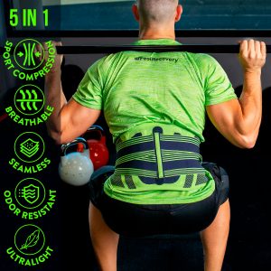
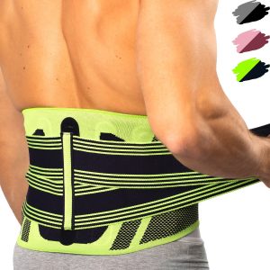
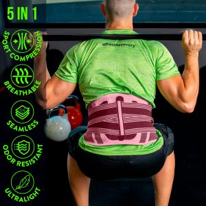
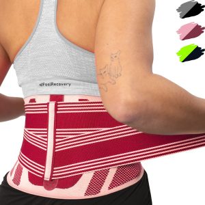
Back Support Belt (Black)
$49.95 -
Back Support Belt (Green)
$49.95 -
Back Support Belt (Pink)
$49.95 -
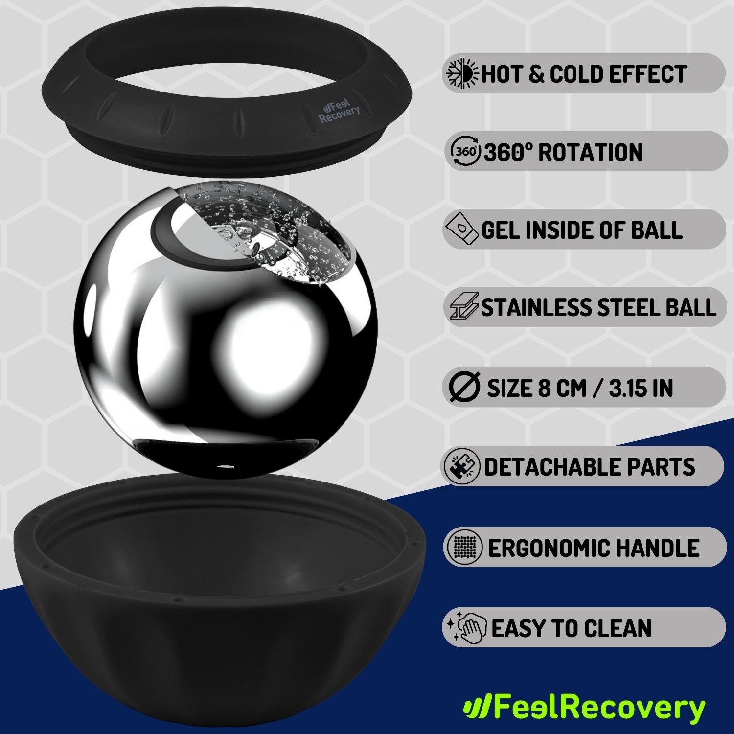
Ice Massage Roller Ball (Black)
$39.95 -
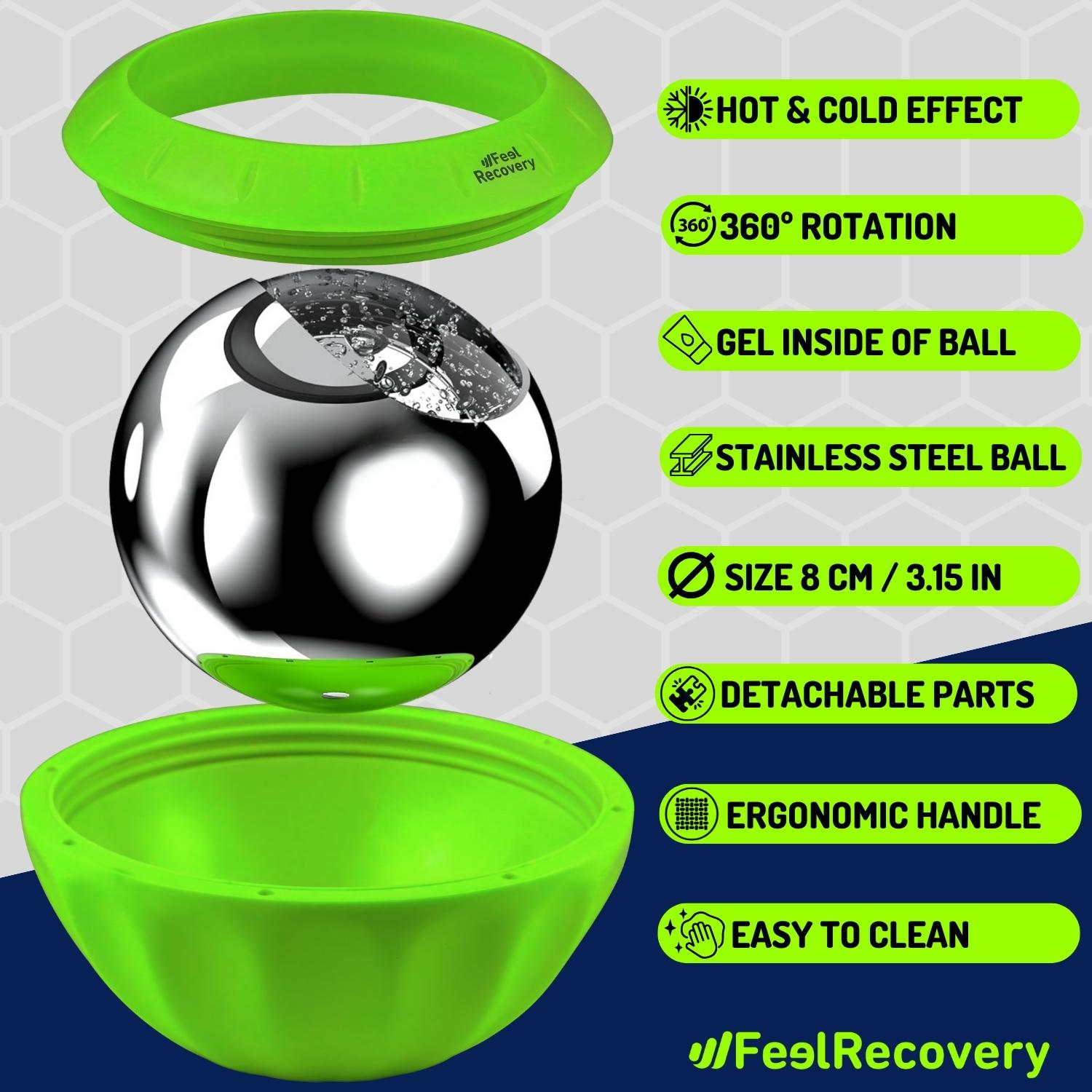
Ice Massage Roller Ball (Green)
$39.95 -
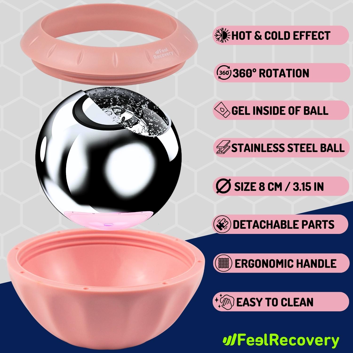
Ice Massage Roller Ball (Pink)
$39.95 -
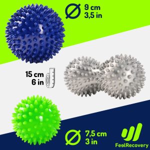
Pack 3 Spiky Massage Balls
$24.95 -
Reusable Gel Ice Packs for Shoulder & Back with Compression Band
$24.95 -
Sacroiliac Support Belt (Black)
$29.95 -
Sacroiliac Support Belt (Green)
$29.95 -
Sacroiliac Support Belt (Pink)
$29.95 -
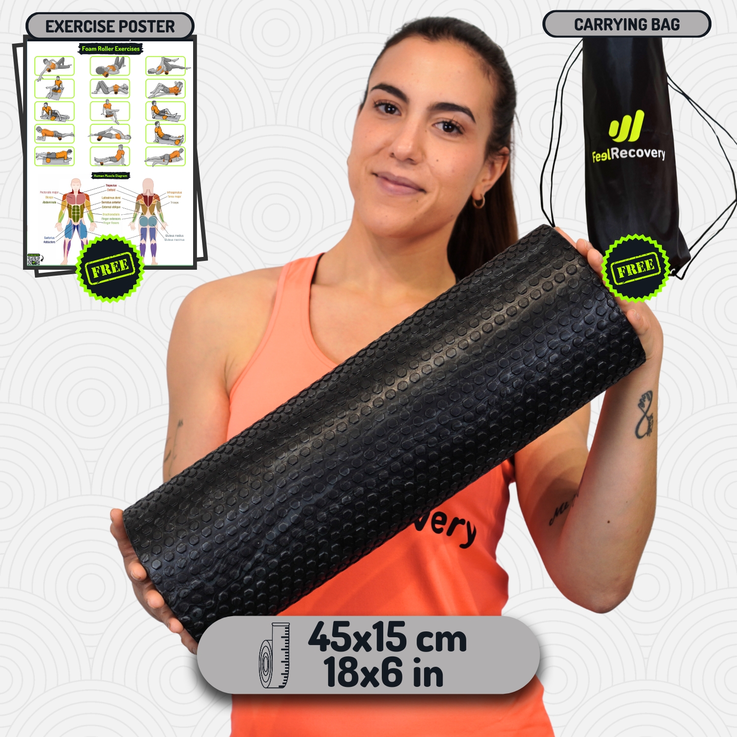
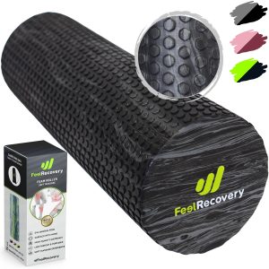
Soft Density Foam Roller for Recovery (Black)
$39.95 -
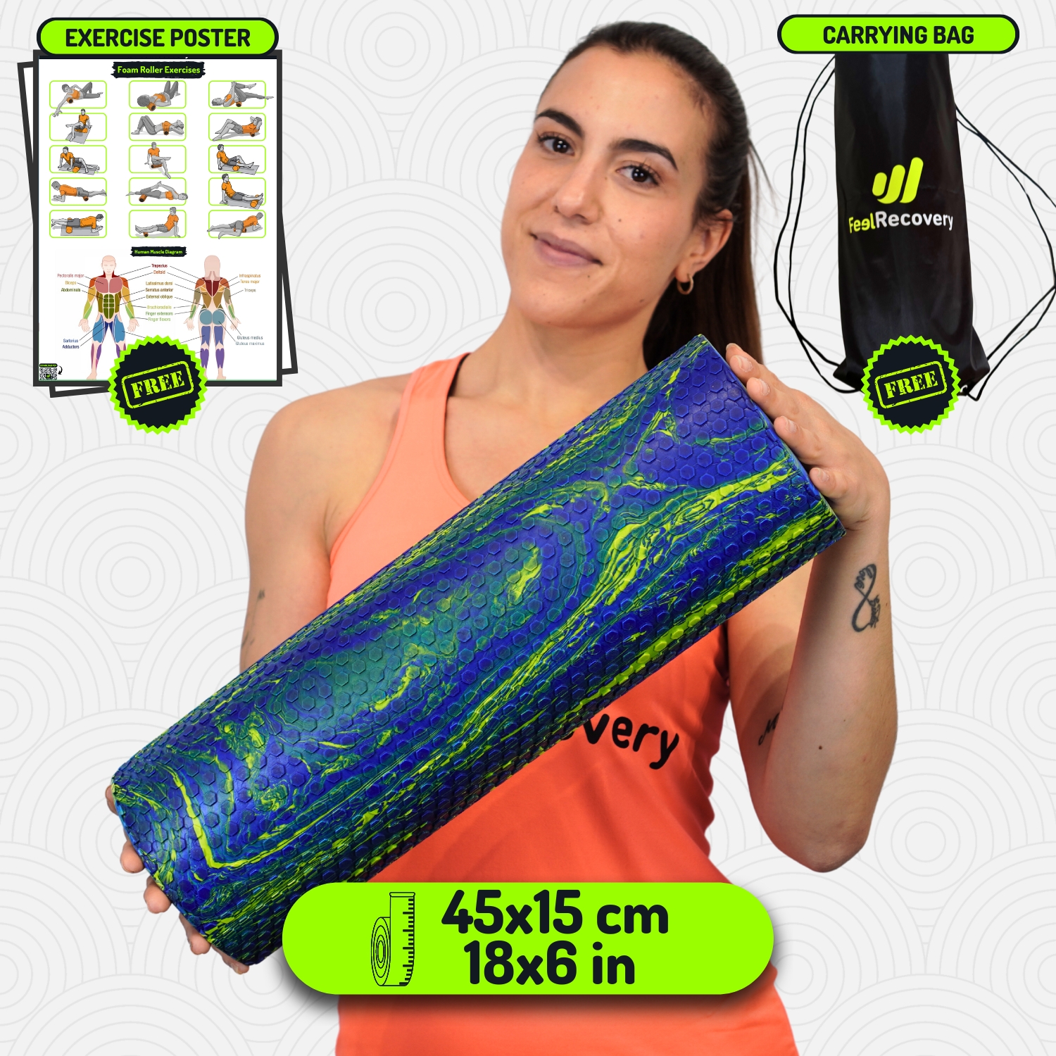
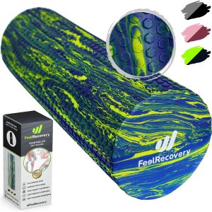
Soft Density Foam Roller for Recovery (Green)
$39.95 -
Soft Density Foam Roller for Recovery (Pink)
$39.95


Biomechanics of the hip joint
Thanks to the ligaments, muscles and bones in the hip, it is possible for the human body to perform different joint movements.
These are discussed below
- Extension: When the leg is placed behind the hip line, the gluteus and hamstrings open 20 to 30°, depending on the flexion of the knee. This movement is limited by the iliofemoral ligament.
- Flexion: With this action it is possible to touch the trunk with the front of the thigh. Its amplitude can be from 90 to 120°, taking into account the flexion that occurs in the knee. In this case the muscles that work are the sartorius, rectus abdominis, tensor fascia latae and psoas iliacus.
- Adduction: This consists of moving the leg to the side, without bending the knee and until it reaches hip height (in the case of lying on one leg), parallel to the axis of the body, causing symmetry with the body. The amplitude of this movement does not exceed 30° and the muscles involved are the pectineus, adductors and rectus internus.
- Abduction: Moving the foot forward, without bending the knee, causes the fascia lata, gluteus and piriformis muscles to work. This movement can have an opening that can reach 180° when the person trains so that the pubofemoral and iliofemoral ligaments do not limit the amplitude.
- Internal rotation: Every time there is an inward movement of the foot, this type of biomechanical action occurs in the hip. The muscles involved are the gluteus medius, gluteus minimus and tensor fascia latae. If the leg is considered as the axis, this movement causes a maximum opening of 40°.
- External rotation: This is the opposite movement to the previous point performed by the foot. In this case the amplitude can reach up to 60° due to the extension of the iliofemoral and pubofemoral ligaments. The quadratus cruris, sartorius, geminus superior and inferior, sartorius, obturator externus and obturator internus muscles are involved.
- Circumduction: These are the circular movements that the hip can perform, taking into account the combination of rotations, adduction, abduction, flexion and extension.
Most common hip injuries
There are different types of injuries that occur in the hip due to the biomechanics of this joint. For this reason, we will explain below which are the most common bruises that occur in this area of the body.
Types of hip injuries
The diseases that can be generated in the hip are:
- Osteoarthritis of the hip: This is one of the most common ailments in the elderly. It consists of the deterioration of the sacroiliac and coxofemoral joints due to wear and tear on the bones and cartilage that join this part of the body. It can be caused not only by age, but also by trauma, autoimmune disorders and obesity, among other risk factors.
- Bursitis of the hip: In each of the hip joints there are sacs containing synovial fluid to improve cushioning and gliding of the joint body. When there is an excess of this fluid, the area becomes inflamed due to the enlargement of this sac. This injury is commonly found in sedentary people and in those who put stress on the hip because of the activity they do.
- Hip fractures: A break in the femoral head or neck of this bone is one of the most common injuries that occur in the hip, although it is also feasible to find breaks in the iliac crest. This can result in total or partial inability to move, permanent pain and bone deformity. Advanced age, osteoporosis and trauma are frequent causes of this ailment.
- Tendonitis of the hip: This injury can occur when any of the ligaments of the hip suffer a strain caused by the accumulation of residual molecules in the tendon fibres. This results in inflammation, pain and loss of mobility in the affected area. Bone malformations and inadequate nutrition are some of the causes of this disease.
- Hip joint dislocation: The coccyx-femoral joint is the space that allows leg movements thanks to the union of the femoral head with the acetabulum of the pelvis. These two bones are joined by cartilage and a series of ligaments, which can cut or move, causing the coccyx to separate from the femur.
Sports injuries to the hip
What we have mentioned so far are the injuries that are generated in the hip according to the diseases. What we will look at next are the contusions that occur in this area with different sporting activities in mind.
Have a look:
- Hip injuries in Yoga: Due to the postures practised in Yoga, it is possible that a person tends to suffer muscular injuries in the hip, especially in the buttocks. In addition, the practice of this activity can also cause some tendons to partially or completely rupture. Bone fractures are rare, but the most common is a broken coccyx.
- Sports injuries of the hip in soccer: The contusions suffered by soccer players in the hip are more related to the breakage of the cartilage inside the pelvic cavity, which can lead to osteoarthritis if it is not healed correctly. Muscle strains suffered in the buttocks, quadriceps and lats are the most common. Athletes can also be seen with inflammation in the bursae of the joints.
- Sports hip injuries in golf: The permanent biomechanical movements of golfers cause over-activity in the joints. This can lead to bursitis and tendonitis. It is also common to find people suffering from pelvic strain.
- Running hip sports injuries: This repetitive and demanding exercise on the hip causes tendinopathies, trochanteric bursitis and muscle strains in the anterior and inner hip area. Some athletes often suffer from impingement between the acetabulum and the femoral head. In more extreme cases the development of osteoarthritis is feasible.
Diseases and ailments in the hip

In addition to cancer and aggressive congenital diseases, it is possible for the hip to suffer from some diseases that we have not yet named. Read on to find out what these conditions are.
Osteoporosis
Osteoporosis is a disease that weakens the strength of the bones causing them to become more fragile, which leads to an increased likelihood of hip fractures. There are different causes for this low bone density, the most common being the patient's age and genetic background.
Dislocations
Hip dislocations arise when there is a loosening at the ends of the bones, causing them to lose their normal position. This can be caused by trauma, blows and activities that place stress on the hip joints. In newborns this is called developmental dysplasia.
Osteochondrosis
Lack of blood supply to the cells within the bone matrix, the osteocytes, causes the stiffness of the bones to be reduced. This causes the bone structure to be weakened and it can break more easily.
Displacement of the femoral epiphysis
When the epiphysis, which is located in the femoral head, slips, either due to trauma or congenital malformations, it causes instability in the hip. Its solution requires the use of pins.
How can we relieve hip bone pain through complementary and non-invasive therapies?
It is possible to apply different complementary hip treatments which are non-invasive and help to relieve bone pain. We will show you below what each of these therapies is about.
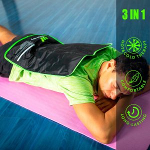
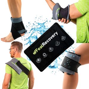
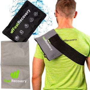
Heat and cold therapy
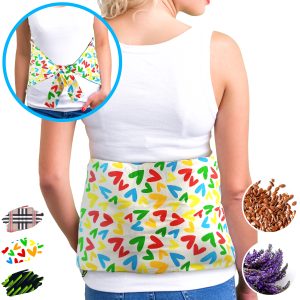
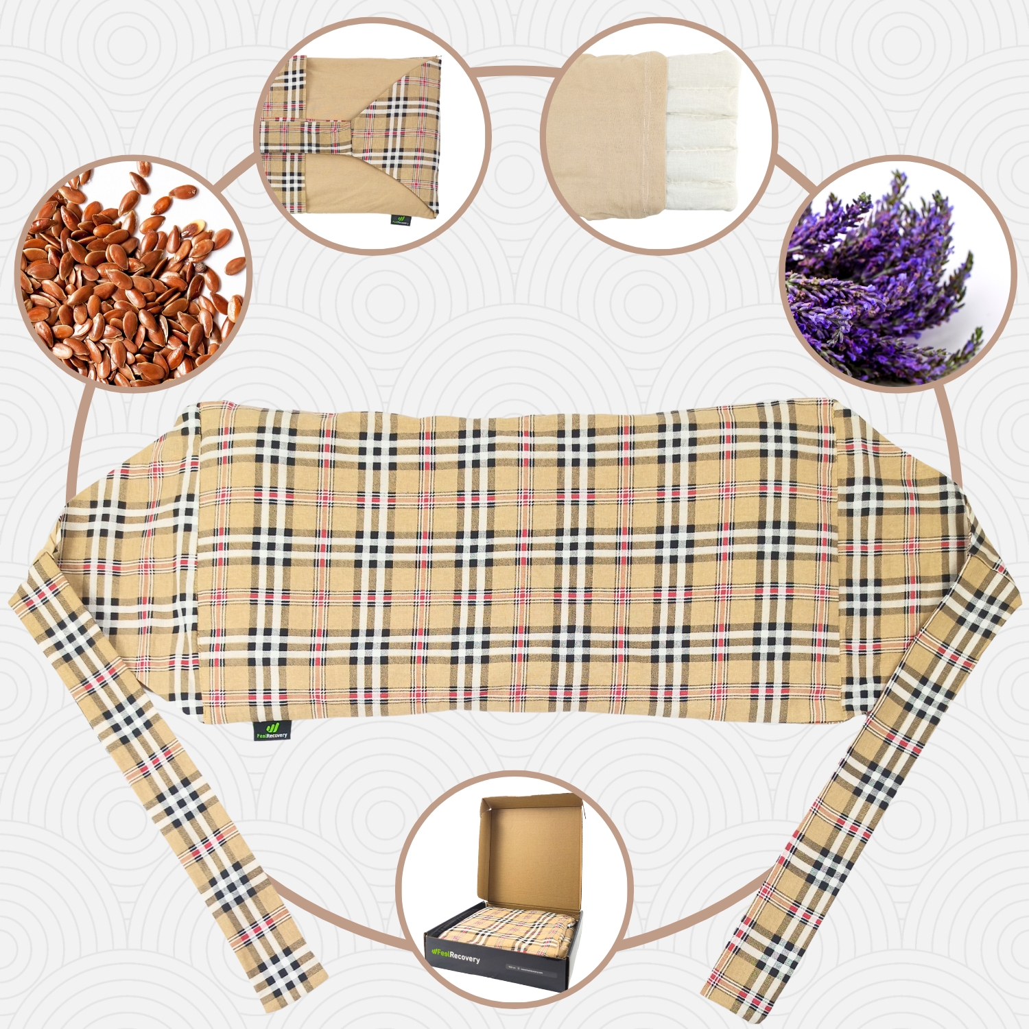
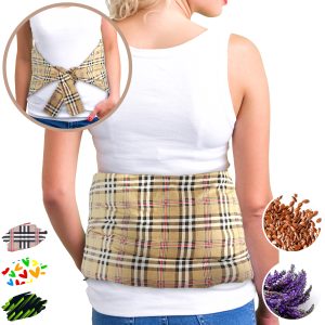
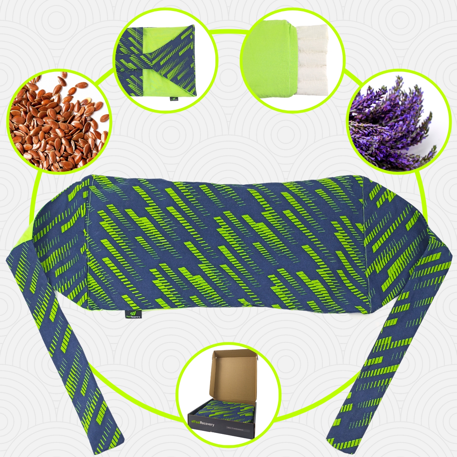
To alternately apply cold and heat to the hip , it is possible to use thermal pillows heating in the microwave and cold gel packs. This provides an anti-inflammatory effect on the muscles, ligaments and tendons located in this area. The application of this therapy should be supervised by the doctor to avoid future injuries, but it is important to note that it cannot exceed 15 to 20 minutes of application in total and should start with the warm temperature.
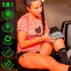
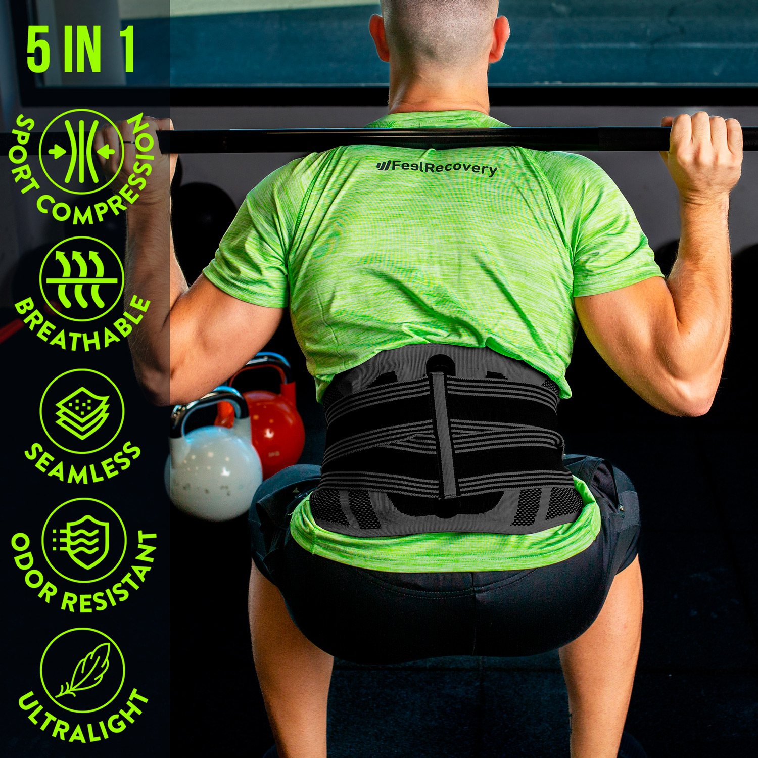
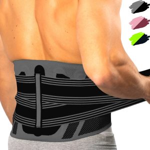
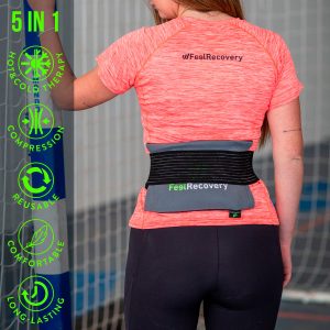
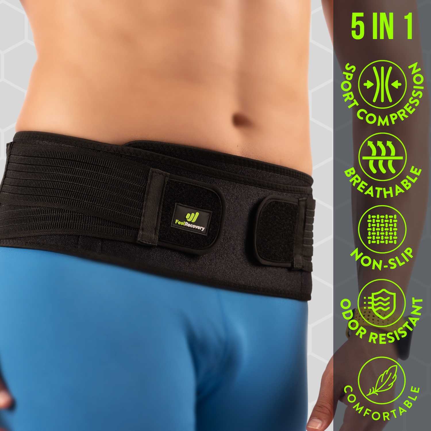
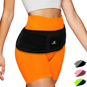
Compression therapy
The hip is one of the most complex joints to recover from. For this reason, lumbar supports, sacroiliac belts and elastic bands are used in non-invasive treatments to hold the hip bones, muscles and tendons in place. This provides a constant and ideal pressure to improve blood circulation and reduce inflammation of the tissues.
Massage therapy
This therapy helps to reduce inflammation and pain in the hip caused by injuries. The aim of this treatment is to restore the patient's mobility as quickly as possible. Different techniques are used for this purpose, the most common being the use of a back massage chair or an electric vibrating ball. Although it is also possible to use physiotherapists to obtain these benefits.
Acupressure therapy
Massage using constant pressure on different areas of the body stimulates the exchange of oxygen between the blood and the soft tissues. This helps to reduce inflammation, restore hip joint movement and reduce the sensation of pain when walking or bending over. This can be done by choosing a physiotherapist or manually using products that fulfil this function, the most commonly used being compression pillows and mats, rollon massagers and hand rollers.
Thermotherapy
The application of heat to the hip is beneficial in stimulating blood flow to the area and reducing the symptoms of the injury. To use thermotherapy it is necessary to use products that generate heat, which can be used personally or by a professional. Among these products are thermal pillows and packs with hot gels, which are applied directly to the affected area. It should be borne in mind that the sessions cannot exceed 15 minutes and can be applied 2 to 3 times a day to avoid more serious risks.
Cryotherapy
Cryotherapy helps to reduce swelling of the hip tissues and improve blood flow to reduce pain in this area. Your doctor may recommend immersion in cold chambers or the use of non-invasive products to help get the benefits of cold. Items that are applied directly to the affected area may include cold gel balls or multipurpose packs with special liquids.
Electrical muscle stimulation (EMS)
Muscle electrostimulation, or EMS, is a therapy that consists of stimulating muscle contractions through the use of electricity, so as to achieve an effect of activity and hypertrophy as in the gym, but without the need to go to any sports center. This means that you can put your muscles to work without leaving home.
Electrotherapy
This is a technique that seeks relief from pain and some physical ailments through the application of electrical and electromagnetic energy, among other variants, through the skin with the use of conductive pads called electrodes. It is a very safe type of therapy and must be applied by a physical therapist specialized in the manipulation of electricity to treat some kinds of ailments.
Myofascial release therapy
Also known as myofascial induction, this therapy consists of the application of manual massage to treat the shortening and tension generated in the myofascial tissue that connects the muscles to the bones and nerves. For this purpose, various massage techniques are used that focus on the so-called trigger points.
Percussion Massage Therapy
Vibration or percussion massages are precise, rhythmic and energetic strokes on the body to achieve relief from some annoying symptoms when muscle fibers are tightened, often by a high workload on them and that has left trigger points in the muscle fibers.
R.I.C.E Therapy
The R.I.C.E. therapy is the first and simplest of the treatment protocols for minor injuries. It appears in the sports field to deal with accidents involving acute injuries. For many years, it has been considered the most suitable for its speed and results.
Trigger points therapy
Myofascial pain points or trigger points are knots that are created in the deeper muscle tissues, causing intense pain. The pain does not always manifest itself right in the area where the point develops, but rather this pain is referred to nearby areas that seemingly do not appear to be related. In fact, it is estimated that more than 80% of the pain they cause manifests in other parts of the body.
Other effective alternative therapies
Other non-invasive treatments that we have not mentioned so far can also be used to treat the hip.
Read on and you will find out what they are all about:
- Natural remedies with herbal remedies: Although the application of herbal baths on the hip is complicated, a herbal tea with herbs containing anti-inflammatory and sedative properties is used instead. In this case, boldo, chamomile, linden and willow bark are used.
- Acupuncture: The symptoms of hip injuries can be treated by means of this Chinese medicine, which consists of strategically inserting special needles. This stimulates internal heat in the musculature and improves the nervous system to help the patient's general condition.
- Kinesiotherapy: If there are bone malformations, muscle contusions or tendinopathies in the hip, it is possible to treat all these symptoms by means of exercises and movements carried out by the kinesiotherapist. In this way it is possible to improve the patient's quality of life.
- Aromatherapy: This non-invasive technique consists of the patient inhaling a fragrance that stimulates psychological balance by means of vapours, particles suspended in the air or by means of soaked cloths. This is possible thanks to the mixture of different aromas that help the patient to relax when suffering pain.
References
- Ng, K. G., Jeffers, J. R., & Beaulé, P. E. (2019). Hip joint capsular anatomy, mechanics, and surgical management. The Journal of Bone and Joint Surgery. American Volume, 101(23), 2141. https://www.ncbi.nlm.nih.gov/pmc/articles/PMC7406151/
- Harty, M. (1984). The anatomy of the hip joint. In Surgery of the hip joint (pp. 45-74). Springer, New York, NY. https://link.springer.com/chapter/10.1007/978-1-4612-5224-5_3
- Byrne, D. P., Mulhall, K. J., & Baker, J. F. (2010). Anatomy & biomechanics of the hip. The open sports medicine Journal, 4(1). https://benthamopen.com/ABSTRACT/TOSMJ-4-51
- McKibbin, B. (1970). Anatomical factors in the stability of the hip joint in the newborn. The Journal of bone and joint surgery. British volume, 52(1), 148-159. https://online.boneandjoint.org.uk/doi/abs/10.1302/0301-620x.52b1.148
- Paluska, S. A. (2005). An overview of hip injuries in running. Sports medicine, 35(11), 991-1014. https://link.springer.com/article/10.2165/00007256-200535110-00005
- Lynch, T. S., Bedi, A., & Larson, C. M. (2017). Athletic hip injuries. JAAOS-Journal of the American Academy of Orthopaedic Surgeons, 25(4), 269-279. https://journals.lww.com/jaaos/Abstract/2017/04000/Athletic_Hip_Injuries.3.aspx
- Boyd, K. T., Peirce, N. S., & Batt, M. E. (1997). Common hip injuries in sport. Sports Medicine, 24(4), 273-288. https://link.springer.com/article/10.2165/00007256-199724040-00005
- Kaufer, H. (1980). Mechanics of the treatment of hip injuries. Clinical Orthopaedics and Related Research®, 146, 53-61. https://journals.lww.com/clinorthop/Citation/1980/01000/Mechanics_of_the_Treatment_of_Hip_Injuries.8.aspx
- Larson, C. M., Swaringen, J., & Morrison, G. (2005). A review of hip arthroscopy and its role in the management of adult hip pain. The Iowa Orthopaedic Journal, 25, 172. https://www.ncbi.nlm.nih.gov/pmc/articles/PMC1888790/
- Bedi, A., Dolan, M., Leunig, M., & Kelly, B. T. (2011). Static and dynamic mechanical causes of hip pain. Arthroscopy: The Journal of Arthroscopic & Related Surgery, 27(2), 235-251. https://www.sciencedirect.com/science/article/abs/pii/S0749806310007267