- What is a shoulder fracture and how is it diagnosed?
- What are the types of shoulder bone fractures there are?
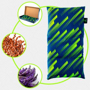
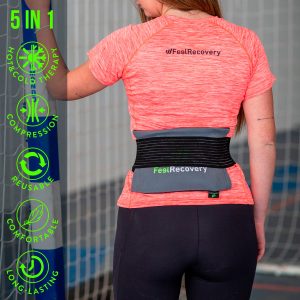
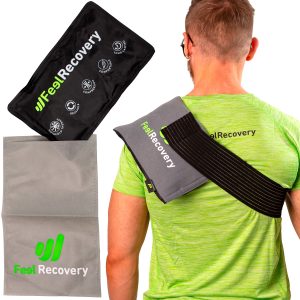
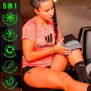
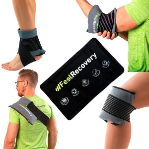
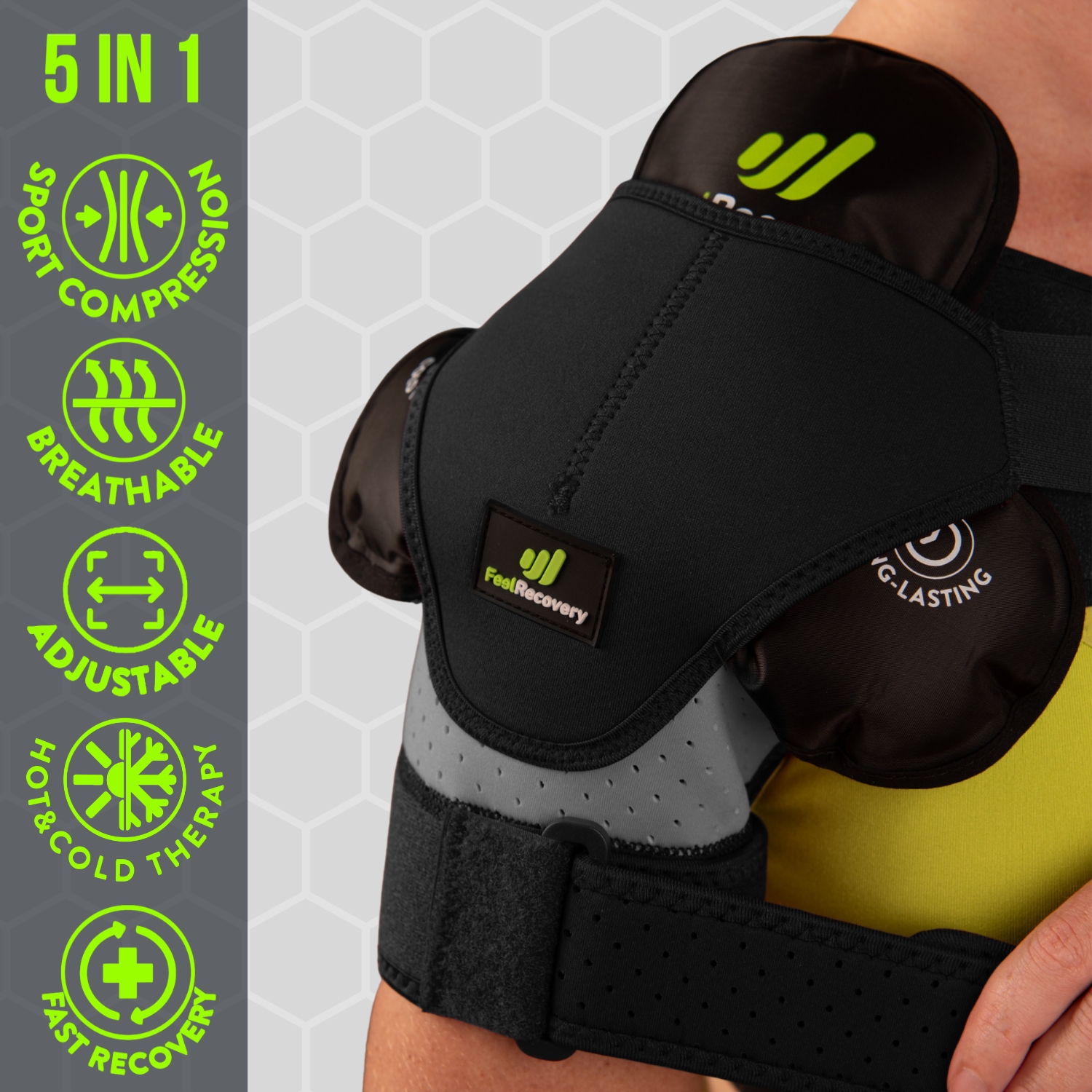
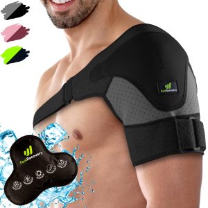
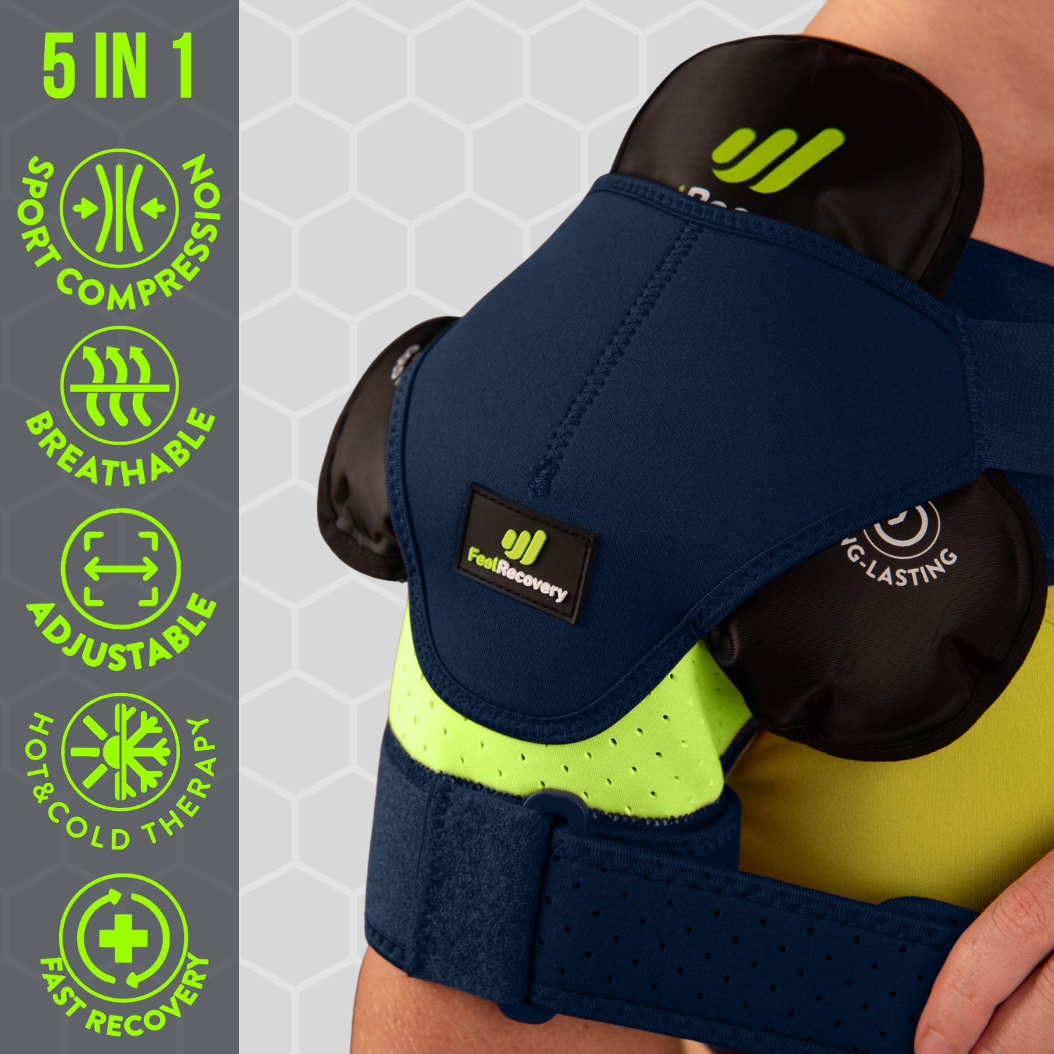
- Best products for shoulder fractures
- What are the causes and risk factors for shoulder fractures?
- First aid care for a shoulder fracture
- The most indicated treatments for clavicle and shoulder fractures
- Rehabilitation after a shoulder bone fracture
- Prevention methods to avoid broken shoulder bones
The shoulder is the joint that connects the arm to the rest of the body and is made up of three bones: the humerus, the scapula and the clavicle. As a result, it is one of the areas of the body most prone to bone fractures.
Shoulder fractures are caused by a crack or break in one of the bones that make up the shoulder joint. As they are injuries of considerable seriousness, it is worth knowing more about what they are, how they are diagnosed, what types are involved, what their causes are, how they should be treated and what methods of prevention exist for this pathology.
What is a shoulder fracture and how is it diagnosed?
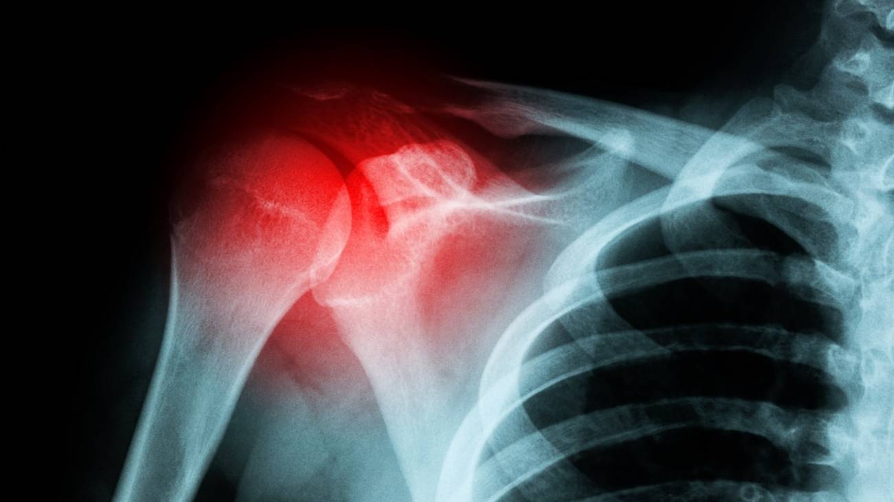
A shoulder fracture is defined as a break or fracture that occurs in the scapula, clavicle and/or proximal humerus. It is also known as a "broken shoulder" and is caused by a crack that directly affects the upper arm bone around the glenohumeral joint. Generally speaking, a fracture corresponds to the loss of continuity of a bone fragment.
The patient manifests a symptomatological picture typical of this type of injury, from which it is possible to identify the broken bone in the upper arm.
The main signs and symptoms that help to detect a shoulder fracture are
- Severe pain in the area of the fracture.
- Swelling or inflammation of the shoulder
- Deformity under the skin
- Skin discolouration or bruising around the break.
- Difficulty or inability to move and lift the arm.
- Extreme tenderness in the area and crunching in the area.
Whendiagnosing a shoulder fracture, medical specialists focus on evaluating the symptoms presented by the patient, in addition to this they opt to carry out a physical study of the fractured region in which the clavicle and its surroundings are examined.
A diagnostic imaging test to visualise the condition of the broken bone is much more accurate and the most common tests are:
- X-rays: In most cases, shoulder fractures can be pinpointed through an x-ray examination. In these, the technician taking the test will need to point to the fracture site from several angles in order to obtain as much detail as possible. In these cases, it is essential that the impacted portion of the bone does not overlap noticeably.
- CT scans: These are imaging tests similar to X-rays in that they also gather X-ray images from multiple angles to externalise the state of the broken bone. However, beyond this, the focus is on combining various techniques to achieve cross-sectional or three-dimensional images of the internal structures of the body. This allows the bones and surrounding soft tissues to be detailed for a more accurate diagnosis.
- Magnetic resonance imaging (MRI): These are more advanced scans that use radio waves along with a magnetic field to make much more detailed images of the ligaments that hold joints and bones together. In effect, the results will allow examination of the break in the bone and also the condition of the ligaments to rule out whether they have been impacted by the injury.
What are the types of shoulder bone fractures there are?

We highlight the classification of this condition according to the bone that has been broken on impact:
- Fracture of the clavicle: This is based on a fracture of the bone that connects the sternum and the shoulder (i.e. the clavicle) or the bone that connects the upper part of the sternum to the shoulder blade. Thus, since the clavicle acts as a stabiliser between the thorax and the shoulder, it stands out as a fracture that should receive immediate medical attention. It is common in children and young adults.
- Fracture of the scapula: Also called a "scapular fracture", this refers to a break that occurs directly in the shoulder blade or scapula; this being the bony portion that connects the collarbone to the upper arm bone. Consequently, if such an injury occurs, the clavicle, arm bones, lungs and even the thorax may also be affected.
- Fracture of the humerus: This refers to a type of break that breaks the humerus or the longest bone of the upper extremity (the upper arm). This is an injury that, in turn, can be segmented according to the region fractured (the lower end, the shaft, or the upper end). Thus, in addition to the common symptoms of a shoulder fracture, it can also lead to deformity of the entire arm and wrist. It usually occurs in the elderly and young.
When a shoulder fracture is diagnosed, it is also essential to point out which pattern has been triggered as follows:
- Non-displaced fracture: This is a bone fracture in which the bone fragments remain in their anatomical location. This means that they do not move from their usual place.
- Displaced fracture: This is a condition in which the bone portions are displaced and/or move away from their regular anatomical location and, as a consequence, exhibit a misalignment. In this case, the degree of displacement and the outline of the fracture must be determined (this alters the natural position of the humerus to a greater or lesser extent).
- Open fracture: This is known as an "open fracture" in which the skin overlying the fracture site is also injured. In other words, the bone fragments break the skin and are exposed to the environment, so that it is possible to see inner tissues and even the fractured bone. This results in an open wound and external bleeding.
- Closed fracture: Unlike the previous case, this is a break that only affects the impacted bone. Therefore, there is no injury to the skin, no open wound and no external haemorrhage. However, internal soft tissue injury and internal bleeding should not be ruled out.
- Simple fracture: As the name suggests, this is a simple bone fracture in which the fractured bone is only splintered in one part.
- Comminuted fracture: When the bone breaks and generates small bone fragments that break off, a comminuted fracture is detected. This means that it is distinguished by the fact that the bone breaks into more than two fragments and is therefore unstable.
- Spiroid fracture: This refers to an injury that triggers a spiral-shaped bone fracture trace. It is almost always caused by a twisting force on the joint.
- Oblique fracture: This is a type of fracture that has an angular pattern, i.e. it causes a break at an angle. This can be with or without displacement.
Best products for shoulder fractures
Bestseller
-

Acupressure Mat and Pillow (Black/Gray)
£44,95 -
Acupressure Mat and Pillow (Green/Navy)
£44,95 -
Acupressure Mat and Pillow (Pink/Bordeaux)
£44,95 -
Acupressure Pillow (Black/Gray)
£21,52 -
Acupressure Pillow (Green/Navy)
£21,52 -
Acupressure Pillow (Pink/Bordeaux)
£21,52 -
Electric Muscle Massager Gun
£129,31 -
Electric Neck & Shoulders Massager
£25,83 -

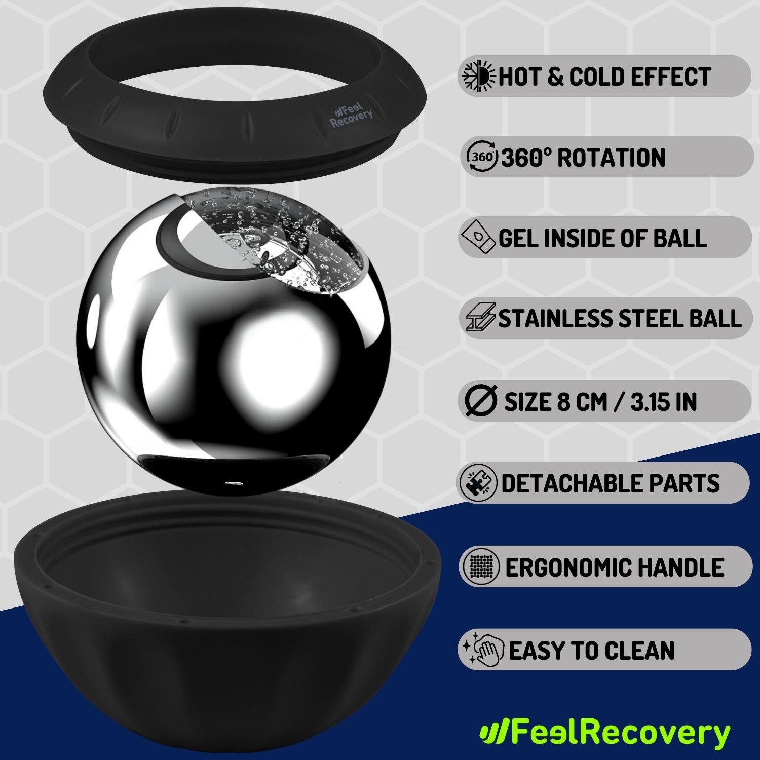
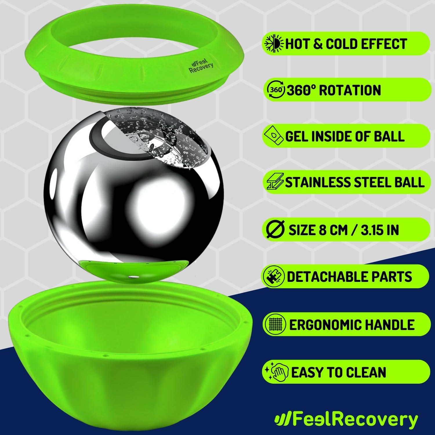
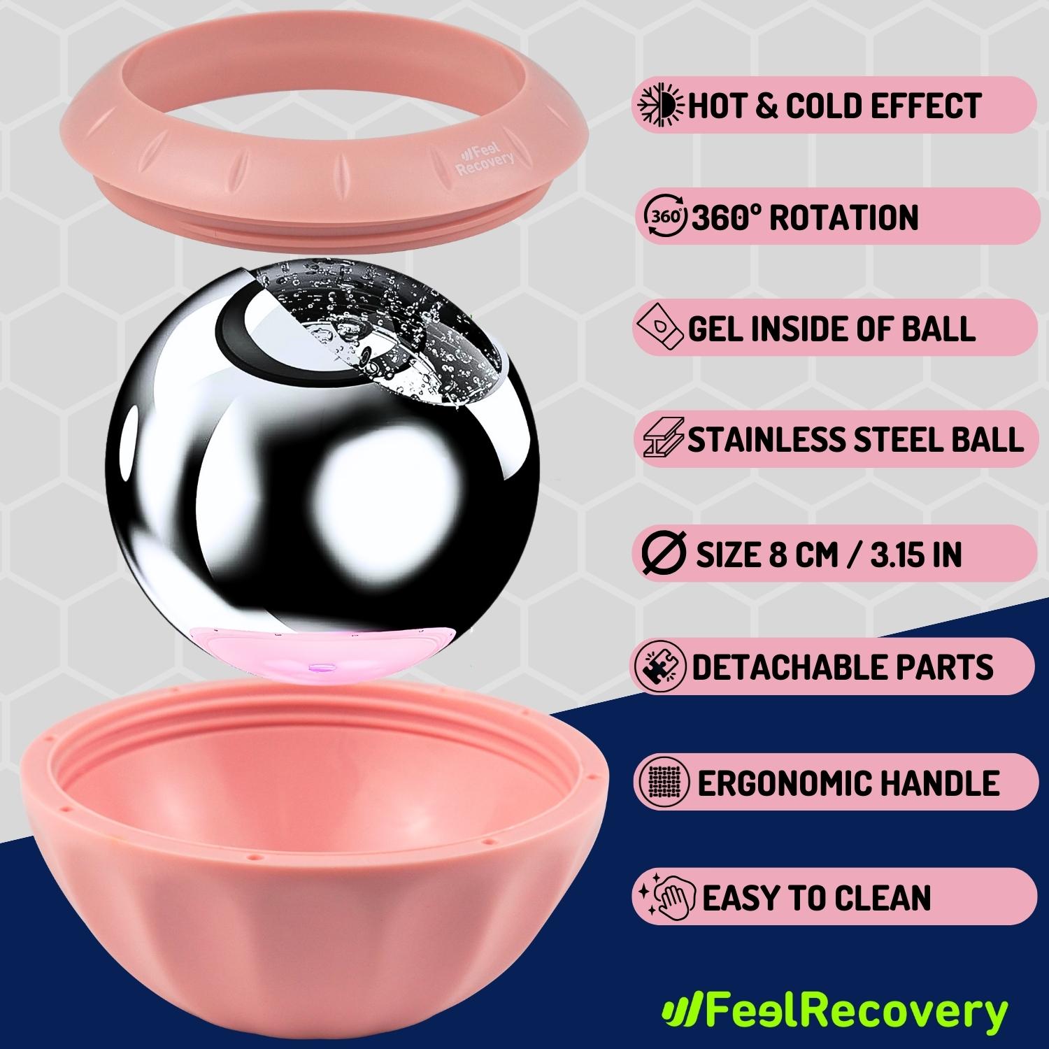
Ice Massage Roller Ball (Black)
£34,95 -
Ice Massage Roller Ball (Green)
£34,95 -
Ice Massage Roller Ball (Pink)
£34,95 -
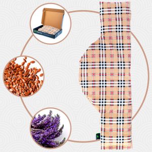
Microwave Wheat Bag for Neck & Shoulder Pain Relief (Hearts)
£21,50 -
Microwave Wheat Bag for Neck & Shoulder Pain Relief (Oxford)
£21,50 -
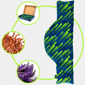
Microwave Wheat Bag for Neck & Shoulder Pain Relief (Sport)
£21,50 -
Microwave Wheat Bag for Neck Pain Relief (Hearts)
£17,50 -
Microwave Wheat Bag for Neck Pain Relief (Oxford)
£17,50 -
Microwave Wheat Bag for Neck Pain Relief (Sport)
£17,50 -
Microwaveable Wheat Bag for Pain Relief (Hearts)
£17,50 -
Microwaveable Wheat Bag for Pain Relief (Oxford)
£17,50 -
Microwaveable Wheat Bag for Pain Relief (Sport)
£17,50 -
Mini Electric Massager Gun
£43,07 -
Reusable Gel Ice Packs for Shoulder & Back with Compression Band
£17,50 -
Reusable Gel Ice Packs with Compression Band for Sports Injury
£17,50 -
Reusable Hot & Cold Packs for Neck & Shoulders Injuries
£21,95 -
Shoulder Support Brace (Black)
£21,95 -
Shoulder Support Brace (Green)
£21,95 -
Shoulder Support Brace (Pink)
£21,95 -
Trigger Point Massage Stick (Black)
£12,95 -
Trigger Point Massage Stick (Green)
£12,95 -
Trigger Point Massage Stick (Pink)
£12,95


What are the causes and risk factors for shoulder fractures?
The origins of a shoulder fracture can vary. Because, by nature, such injuries are the result of numerous risk factors and eventualities that suddenly break one or more bones in the upper arm.
Here we highlight the main risk factors for a broken bone in this area:
- Falls: Especially if the individual falls directly on the shoulder, the event occurs with the arm outstretched or the hand outstretched.
- Direct blows and trauma to the shoulder or the humerus (upper arm) also tend to trigger bone fractures in this region of the body.
- Automobile, motorbike and bicycle accidents generate high impacts that cause severe injuries, including fractures to the shoulder bones.
- Sports injuries that spread sudden forces applied to the shoulder, whether on a track, court or playing field, are other major causes.
- Engaging in physical activity and suddenly increasing its frequency, duration and/or intensity can lead to a break in the shoulder bones due to a stress fracture. Likewise, if you perform movements incorrectly and use contraindicated sports equipment, you are likely to suffer a fracture due to trauma or a fall.
- Conditions that reduce bone density or weaken bones directly, such as osteoporosis, increase the risk of a shoulder fracture.
- Although this type of injury also occurs in young people and children, another contingency factor is age. As the bones become much weaker and more fragile over the years, they are prone to break easily.
- If a person has an unhealthy diet, smokes and drinks alcohol, he or she is associated with a high risk of broken bones. Bad habits lead to bone loss, wear and tear of these structures and even promote the development of osteoporosis (especially smoking).
First aid care for a shoulder fracture
When a patient fractures her shoulder, the first step is to seek trained medical help who can treat the injury appropriately. However, during the time it takes to receive such help, it is essential that both the injured person and others attempting to help them after their accident take certain precautions to prevent the fracture from increasing in severity and causing further harm to the patient.
The best first aid measures to implement in the event of a fracture of a bone fragment of the shoulder are:
- Keep the individual's shoulder immobile: It is essential to avoid any type of mobility of the shoulder and to encourage the patient to leave the joint static. If the patient does any activity or movement, the pathology is likely to worsen and become more severe. By leaving it immobile, it will be possible to limit the pain and discomfort caused by the fracture.
- Leave the broken bone as it is after the impact: It is not advisable to try to align the broken bone or to push it inwards or touch it. This not only increases the pain, but can also graft on the bony portion in a negative way and even trigger an infection in the area (if it is an open fracture where the bone protrudes through the skin).
- Avoid taking any medication without a doctor's prescription: It is possible that, after experiencing the injury, the patient may choose to take an over-the-counter drug in order to lessen the pain and discomfort that persists due to the break in the bone. However, this can be detrimental to their health, due to the side effects that develop and thus stimulate the onset of other pathologies. Apart from that, it is suggested to avoid consuming food or drinking liquid.
- Monitor the patient's vital signs: Because of the impression or symptomatological picture manifested by the person, it is likely that their vital signs are altered. Consequently, it is ideal to monitor the patient's state of health by means of these indicators to corroborate that they are in a stable and conscious condition, as well as to prevent the severity of the pathology. So, as far as possible, it is necessary to take their pulse, measure their temperature, calibrate their respiratory rate, blood pressure and blood pressure, etc. Even prevent them from closing their eyes and falling asleep.
- Try to control their nervousness: Although many injured people remain calm, the truth is that other people go into a state of shock and show a noticeable nervousness due to the impact they have suffered. However, this tends to increase the pain and worsen the condition of the broken bone. Therefore, rescuers should try to help the person to relax in order to promote stability in all respects while obtaining clinical care.
- Apply cold compresses to the shoulder: Naturally, cold has properties from which it provides an analgesic, anti-inflammatory and sedative effect. As a result, it stimulates the reduction of pain, the elimination of inflammation and helps to relax the traumatised area. For this reason, it is recommended to apply cold compresses to the shoulder to soothe the main symptoms of the bone fracture. It is counterproductive to use ice directly on the skin, as it causes burns.
The most indicated treatments for clavicle and shoulder fractures
In general, treatment of a fracture of the shoulder and collarbone will always depend on the number of bone fragments that have broken off at the site, as well as their displacement.
To avoid the possible complications of such a fracture, it is essential to take into account other factors such as the following: fracture pattern, condition of the stabilising structures of the shoulder, level of activity and degree of osteoporosis of the individual.
If the shoulder fracture is non-displaced or only minimally displaced according to Neer's criteria, conservative treatment should be applied. This refers to closed reduction of the injury and relies on immobilisation of the affected limb (i.e. the shoulder) by means of a sling for an appropriate period of time.
Depending on the radiographic tests performed by the health professional, the health professional can stipulate when it is appropriate to remove the immobilising element:
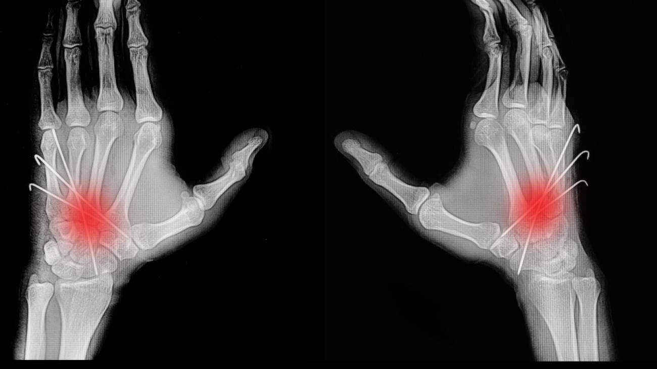
- Closed reduction and percutaneous fixation: For this procedure, closed reduction manoeuvres are used to place the affected bone in its corresponding anatomical position, with the help of the image intensifier and thus, it is anchored by means of needles. As a result, it does not promote damage to the vascularisation of the area and shows less scar tissue formation (which hastens the recovery of the shoulder).
If the fracture in this region of the body shows noticeable displacement and reaches a higher level of severity, surgical intervention is mandatory to replace the injured bone fragment and restore its function. In most cases, screws, pins or metal plates are used to help hold the bones in place while they heal.
These are the procedures associated with shoulder fracture surgery:
- Osteosynthesis plates: In a type of surgery that is usually used when the functional demands of the patient will be high and in fractures of greater complexity. These are plates that are fixed to the bone by means of screws and are responsible for stabilising it throughout the healing process. This requires an open procedure consisting of incisions in the skin and surrounding soft tissue. In this way, it attempts to reduce the tuberosities, restore the length of the bone and restore retroversion to its head, thus supporting the anatomical reduction of the bone fragments.
- Intramedullary nails: Although previously, this type of nail was reserved specifically for diaphyseal fractures of the humerus, nowadays, certain nails are used that maintain a high level of stability in bone injuries of the proximal humerus. In addition, it is considered an optimal solution for simple fractures and those with large metaphyseal extension. Its greatest benefit is that these devices can be implanted in a minimally invasive manner, thereby reducing damage to the vasculature or periosteum.
- Prosthetic replacement: If the fracture in a shoulder bone becomes acute and the lack of blood supply causes degeneration in the area or another complication appears, despite good fixation of the break, it is necessary to resort to a prosthetic replacement. In other words, this type of intervention is usually suggested in complex fractures (three and four fragments) or in articular fractures. Taking into account that there are various types of prostheses that are adapted to the specific case of each patient.
Rehabilitation after a shoulder bone fracture

Once the initial treatment is completed, the patient must undergo rehabilitation treatment to reduce or eliminate the stiffness of the broken shoulder bone. In addition, this recovery phase will aim to improve the motion of the joint, restore muscle strength and increase the flexibility of the shoulder.
Post-treatment involves shoulder rehabilitation exercises that are directed by a trained physiotherapist who, depending on the patient's diagnosis and the treatment they have undergone, will establish a personalised plan to accelerate their recovery process.
Depending on the area of the fracture, the following rehabilitation processes will be carried out:
Clavicle fractures
Rehabilitation during and after immobilisation is distinguished by the following:
- During immobilisation: On the second or third day, the scapula is fixed and small active-passive movements of arm rotation, soft isometric movements of the deltoid, isometric movements of shoulder flexion-extension, retraction and protrusion can begin.
- After immobilisation or following osteosynthesis plate surgery: A sedative management is initiated, followed by a progressive mobilisation process that after being active-assisted, becomes fully active. Thus, the recommended movements are as follows: descent, elevation, retraction, protrusion, progressive muscle toning and joint mobilisation (if there is a lot of stiffness and there is complete bone consolidation).
Fractures of the scapula
The procedure is as follows:
- If immobilised: Under this condition, once the first few days have passed, it is appropriate to start active-assisted movements and in most cases, a home plan is drawn up for the patient that must be followed responsibly.
- After surgical treatment: If an operation is performed, the first step after the operation is passive mobilisation and muscle strengthening.
- After treatment: During the final rehabilitation plan, when the scapula is fully consolidated, it is possible to undertake progressive mobilisation. In addition to this, progressive toning should also be carried out and it is important to avoid muscle contractions during the strengthening phase.
Humerus fractures
The rehabilitation process after a humerus fracture is as follows:
- Immobilisation: If no contraindications are observed, progressive mobilisation of the scapulohumeral type should be carried out. Apart from this, regardless of whether the patient is in a cast or sling, it is important to perform active assisted exercises.
- After immobilisation: Afterwards, to avoid fatigue, it is advisable to use the sling only on an occasional basis. It is valuable to do passive exercises while the pain is controlled (with great care) and to increase the amplitude and strength of the exercises gradually. Early isometric exercises and resisted exercises in functional diagonals are also recommended.
- During the period of total immobilisation: Active mobilisation of the fingers, wrist, elbow and cervical spine is used.
- During the period of relative immobilisation: Isometric exercises that do not intervene in the fracture or produce traction are ideal. As well as painless active-passive mobilisation in small amplitudes and without rotation (abduction, adduction, protrusion and retraction).
- Consolidation period completed: Isometric exercises and isotonic exercises are performed.
Prevention methods to avoid broken shoulder bones
There are different methods of prevention to reduce the risk of suffering this type of fracture, the most common are:
- Have a healthy and balanced diet: Eating a balanced diet will help your body to function optimally. In addition, by making sure you eat foods high in calcium and vitamin D, you can promote the development of bone strength to prevent weak bones.
- Engage in physical activity consistently and progressively: If you exercise your body on a daily basis, in addition to strengthening muscles to prevent sudden falls, you can also increase the amount of bone and stimulate joint health. Keeping in mind that it is important to do it gradually to prevent overtraining from causing injuries that can break down bone fragments.
- Wear appropriate footwear for each occasion: As falls are one of the main causes of these fractures, it is suggested to use shoes that fit your feet perfectly, are non-slip and are considered suitable for each skill to be practised. In addition, keep in mind that it is appropriate to replace athletic shoes periodically (especially if they have irregular wear) and it is not advisable to wear high-heeled shoes excessively.
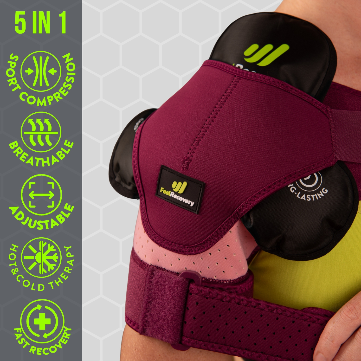
- Make sure you wear protective equipment during sporting activities: If you engage in physical activity (especially if it is high-risk), it is vital to wear protective equipment that protects the area to prevent a fracture in the event of a direct blow. Adjustable shoulder pads or compression bandages are useful to avoid breaking shoulder bones.
- Drive carefully and don't forget to wear your seatbelt: Traffic accidents increase the risk of fractures to the shoulder or any other part of the body, so it is appropriate to drive carefully and wear your seatbelt at all times.
- Don't practice habits that are harmful to your health: If you smoke or drink alcohol frequently, we advise you to stop doing so to protect your well-being. We value the fact that smoking stimulates the manifestation of osteoporosis and alcohol causes bone loss, as well as generating disorientation and thus tends to cause falls or traffic accidents.
- Prevent falls in your home by clearing the floor: It is essential to keep the floor of your home completely clear and conditioned for walking without the risk of tripping or suffering domestic accidents. To do this, the most basic thing to do is to keep it free of clutter, remove loose cables, pick up slippery rugs, leave furniture or decorative objects in their usual place and avoid cleaning the floor using slippery wax.
- In the kitchen, it is suggested to have non-slip mats in the area (especially near the sink) to prevent sudden slips. In addition, it is valuable to keep the floor completely clean with no spilled liquids or food that could cause a fall (both in the kitchen and elsewhere in the house).
- In bathrooms, you can place rubber mats that are non-slip to avoid impacts that affect the shoulder and/or use bath slippers with soles that prevent slipping on wet surfaces. It is also advisable to have bars on the bathroom walls for easy grabbing and, if a person is unable to stand up safely, it is appropriate to use a plastic chair with a backrest in the shower.
- On stairs, consider leaning on handrails whenever going up or down stairs as this helps to prevent falls on stairs. On the other hand, if you have stairs in your home, you can install handrails on each side to implement this recommendation. It is even appropriate to tape bright tape on the steps to differentiate them easily and to keep the area well lit by setting light switches there.
References
- McKoy, B. E., Bensen, C. V., & Hartsock, L. A. (2000). Fractures about the shoulder: conservative management. Orthopedic Clinics of North America, 31(2), 205-216. https://www.sciencedirect.com/science/article/abs/pii/S0030589805701413
- Gallinet, D., Adam, A., Gasse, N., Rochet, S., & Obert, L. (2013). Improvement in shoulder rotation in complex shoulder fractures treated by reverse shoulder arthroplasty. Journal of shoulder and elbow surgery, 22(1), 38-44. https://www.sciencedirect.com/science/article/abs/pii/S1058274612001206
- Herscovici Jr, D. O. L. F. I., Fiennes, A. G., Allgower, M., & Ruedi, T. P. (1992). The floating shoulder: ipsilateral clavicle and scapular neck fractures. The Journal of Bone and Joint Surgery. British volume, 74(3), 362-364. https://online.boneandjoint.org.uk/doi/abs/10.1302/0301-620X.74B3.1587877
- Kokkalis, Z. T., Iliopoulos, I. D., Antoniou, G., Antoniadou, T., Mavrogenis, A. F., & Panagiotopoulos, E. (2017). Posterior shoulder fracture–dislocation: an update with treatment algorithm. European Journal of Orthopaedic Surgery & Traumatology, 27, 285-294. https://link.springer.com/article/10.1007/s00590-016-1840-5
- Murray, A. W., McQuillan, C., Kennon, B., & Gallacher, S. J. (2005). Osteoporosis risk assessment and treatment intervention after hip or shoulder fracture: a comparison of two centres in the United Kingdom. Injury, 36(9), 1080-1084. https://www.sciencedirect.com/science/article/abs/pii/S0020138305001208
- Müller, M. E., Nazarian, S., Koch, P., & Schatzker, J. (2012). The comprehensive classification of fractures of long bones. Springer Science & Business Media. https://books.google.es/books?hl=en&lr=&id=t0CgBQAAQBAJ
- Watson-Jones, R. (1962). Fractures and joint injuries. E. & S. Livingstone. http://117.239.25.194:7000/jspui/bitstream/123456789/1853/7/INDEX.pdf
- Hoppenfeld, S., & Murthy, V. L. (Eds.). (2000). Treatment and rehabilitation of fractures. Lippincott Williams & Wilkins. https://books.google.com/books?hl=en&lr=&id=bxhycwgJUtQC
- Kanis, J. A., Borgstrom, F., De Laet, C., Johansson, H., Johnell, O., Jonsson, B., … & Khaltaev, N. (2005). Assessment of fracture risk. Osteoporosis international, 16, 581-589. https://link.springer.com/article/10.1007/s00198-004-1780-5
- BAILEY, M. (1982). Emergency! First aid for fractures. Nursing2021, 12(11), 72-81. https://journals.lww.com/nursing/Citation/1982/11000/EMERGENCY__FIRST_AID_FOR_FRACTURES.22.aspx