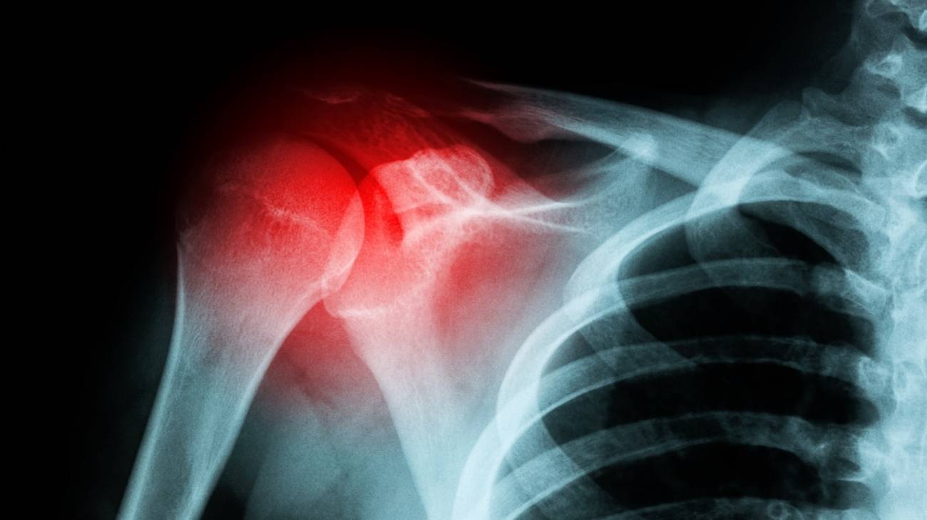
- What is a fracture of the bones of the foot and how is it diagnosed?
- What are the types of foot bone fractures there are?
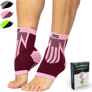
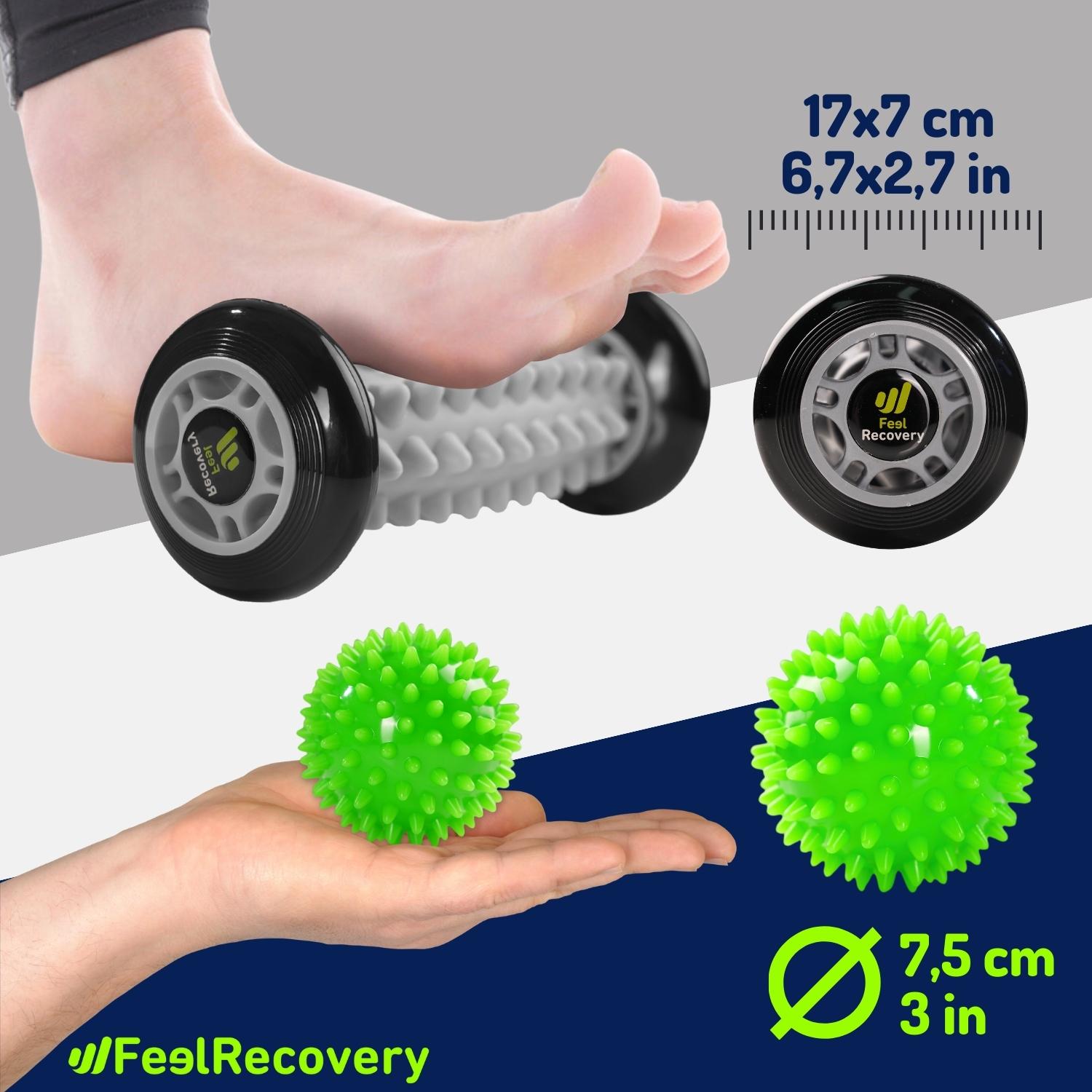
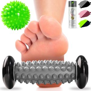
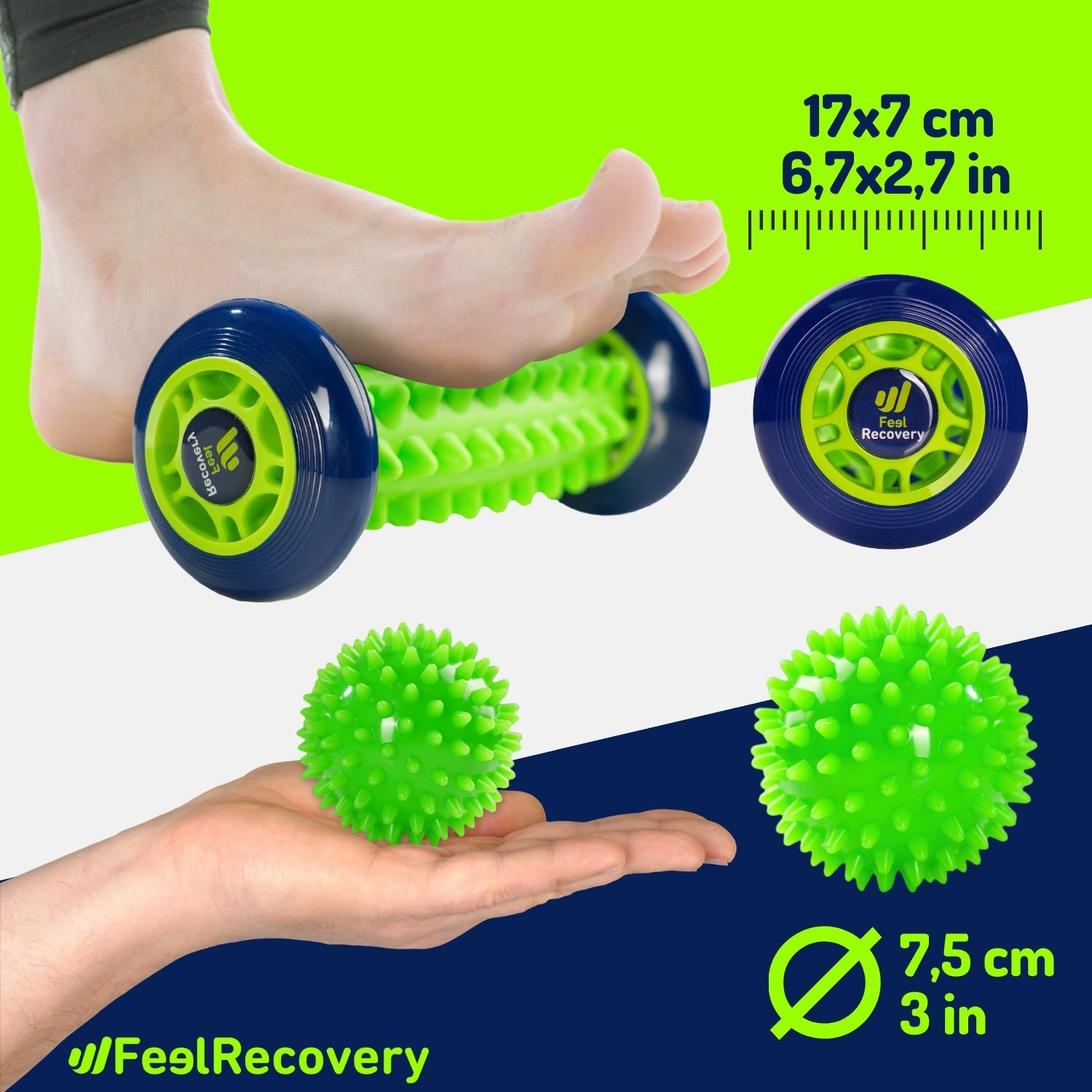
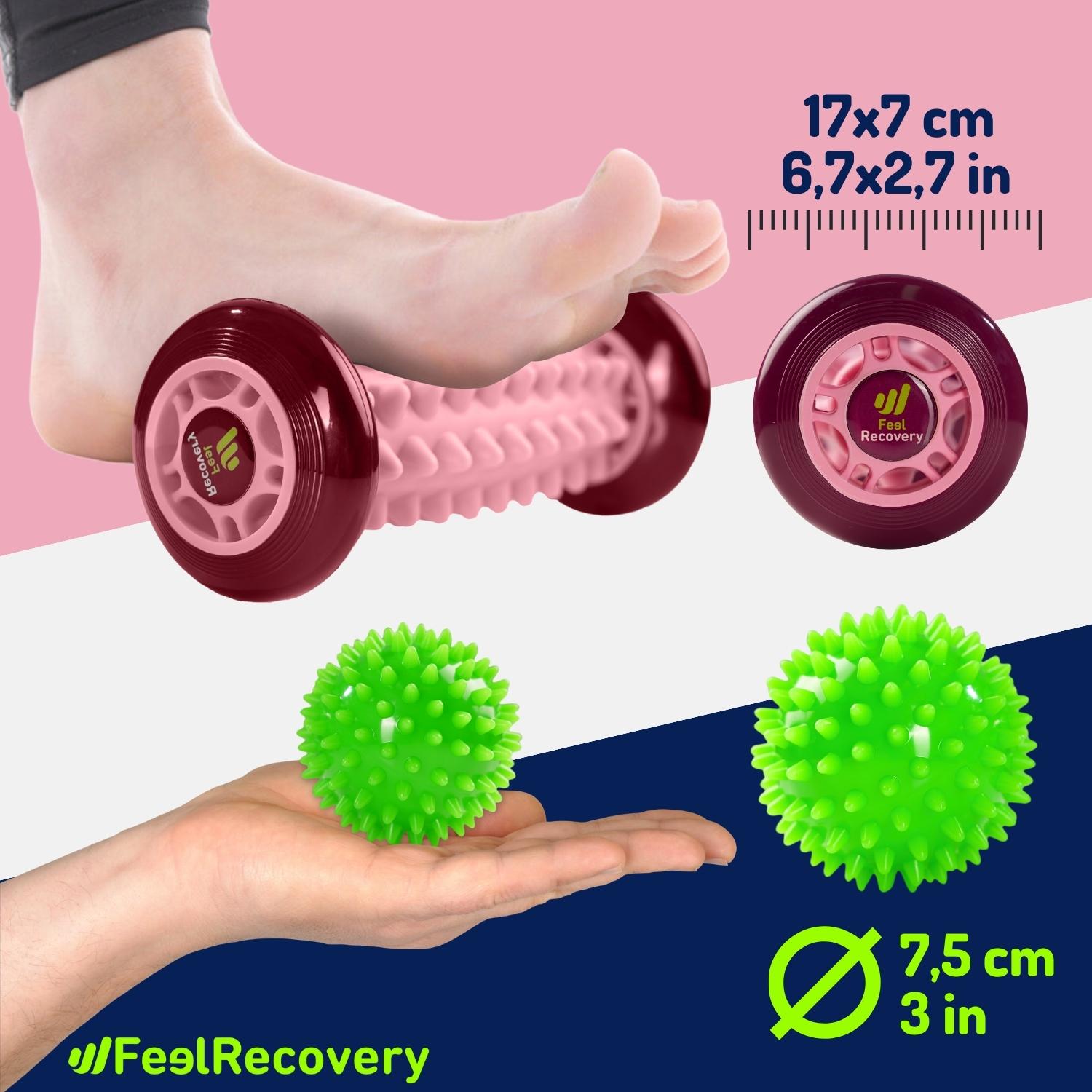
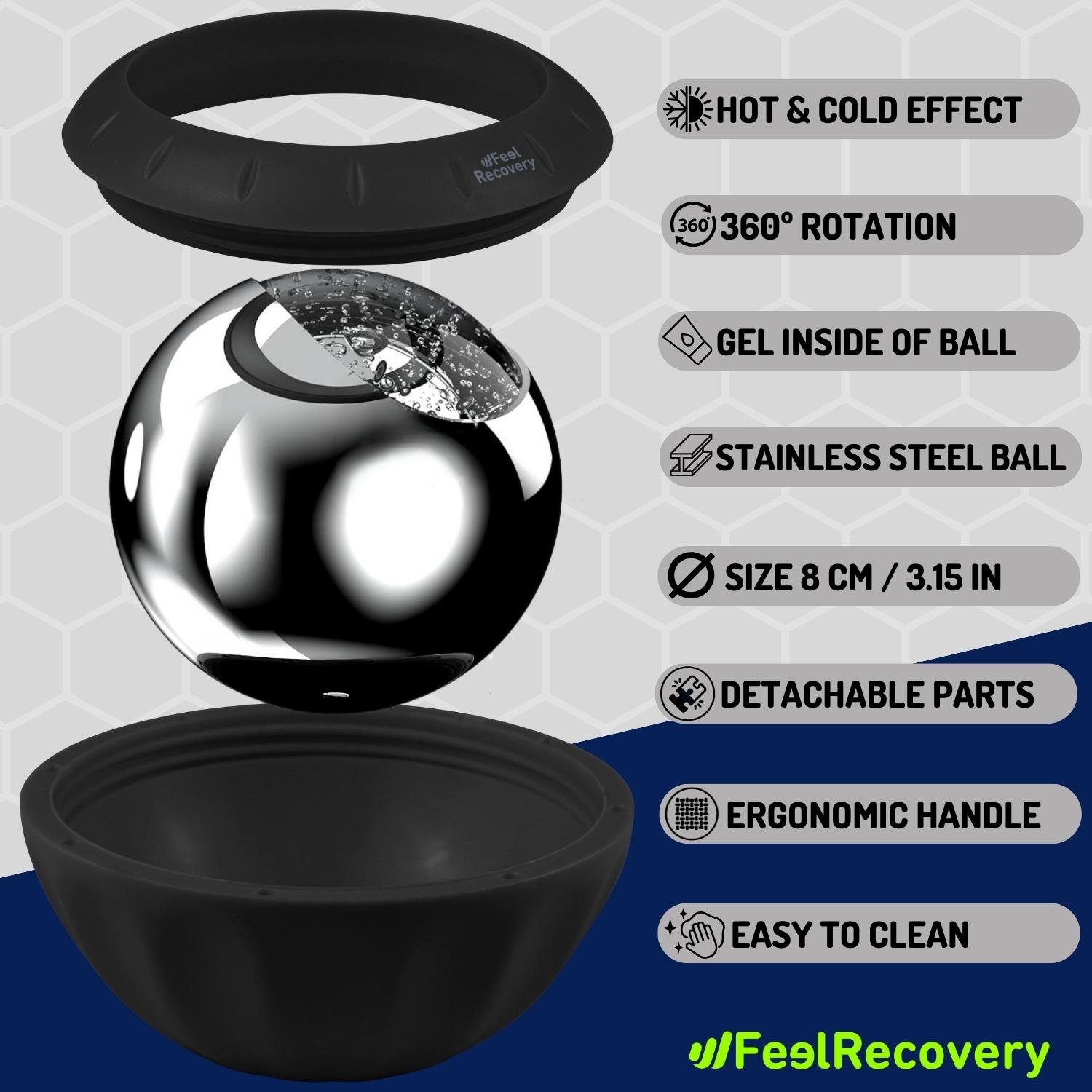
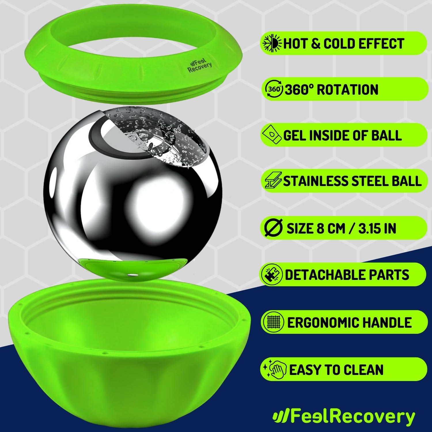
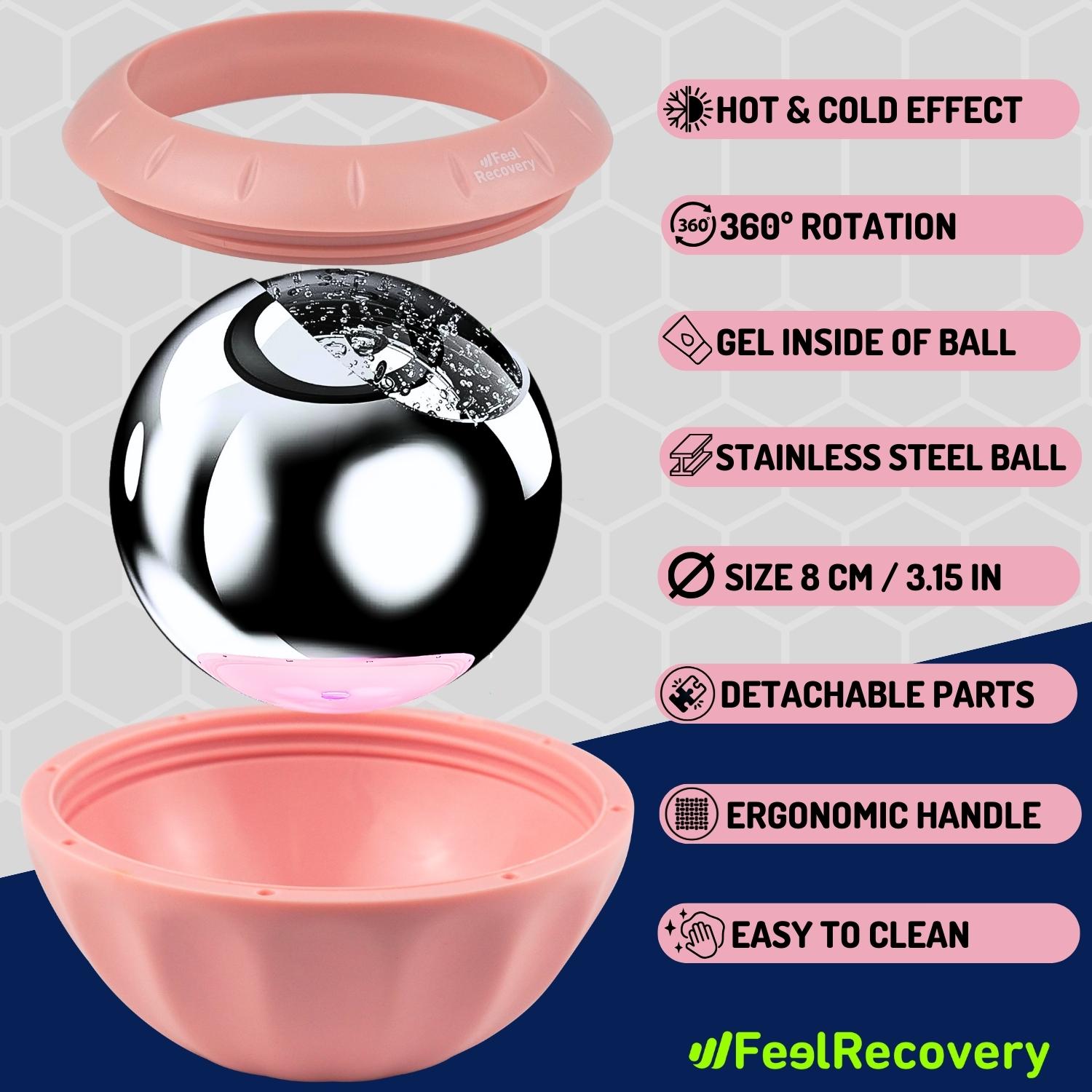
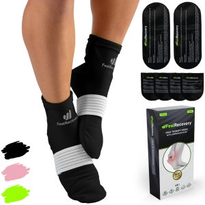
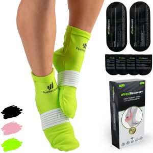
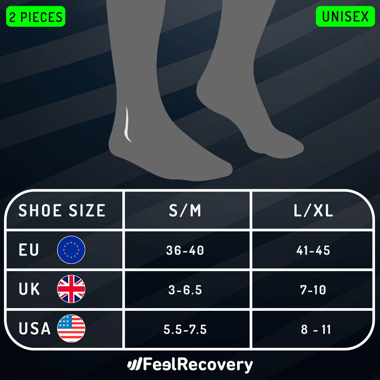
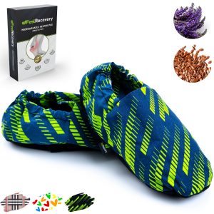
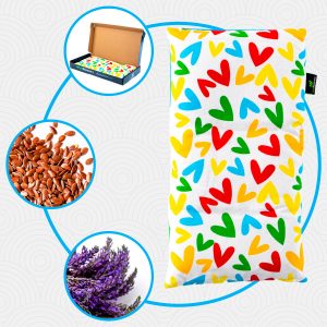
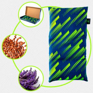
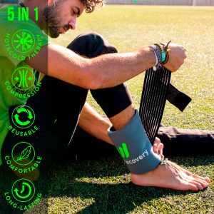
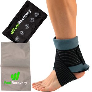
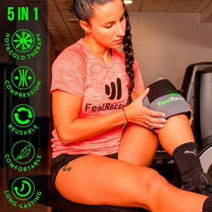
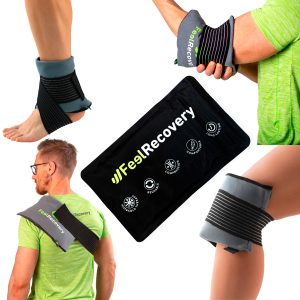
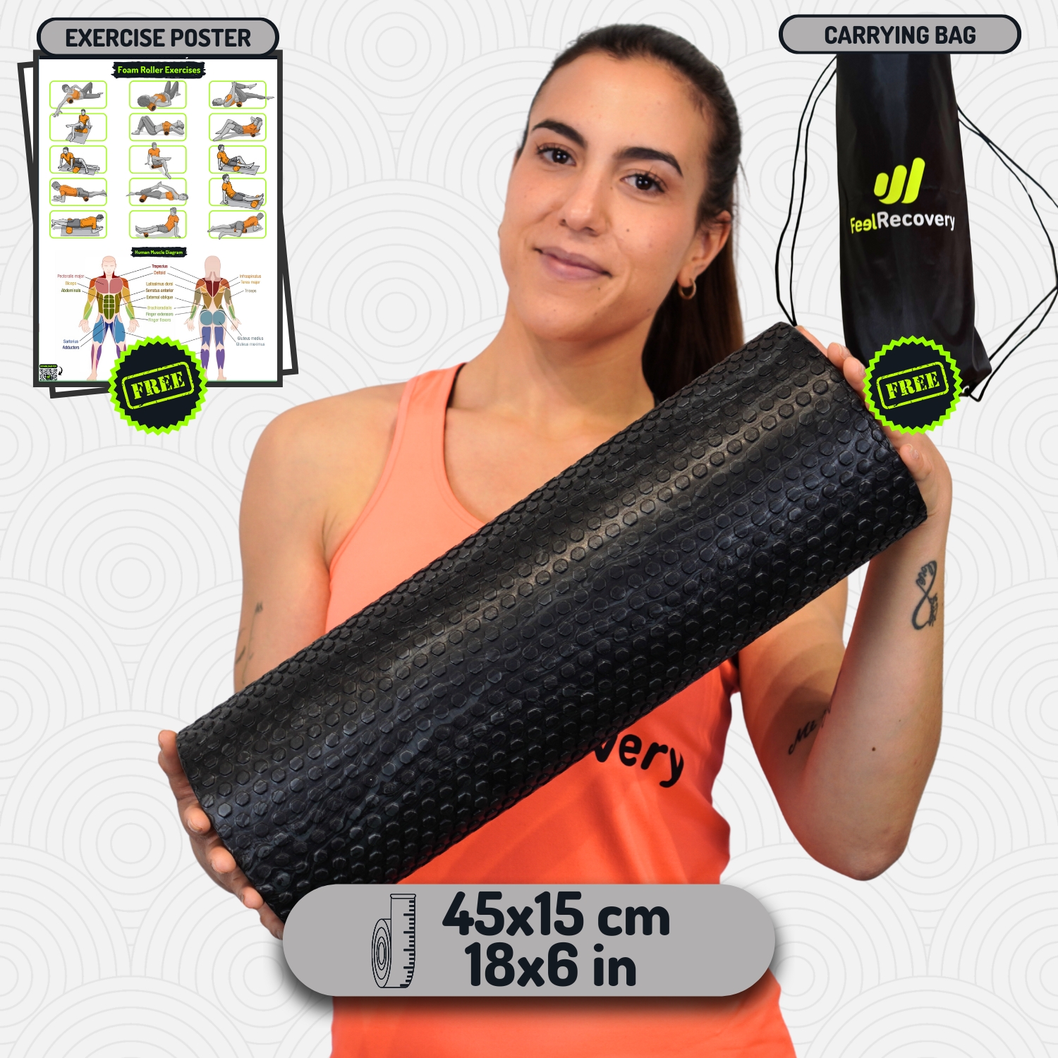
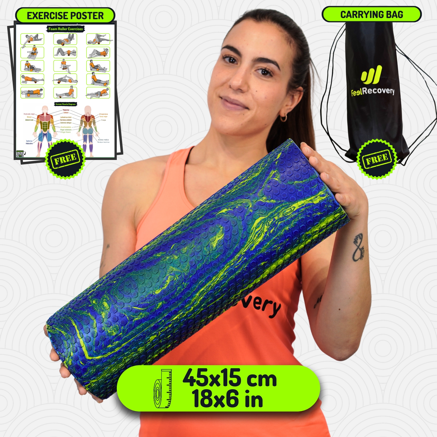
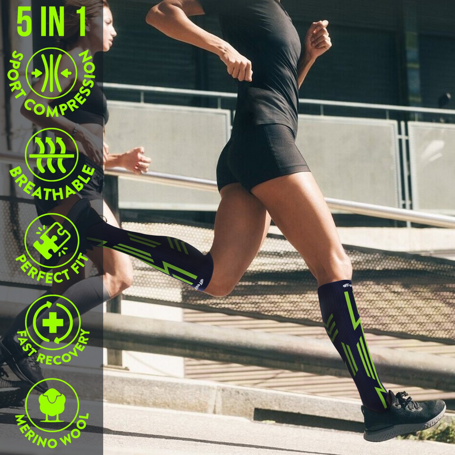
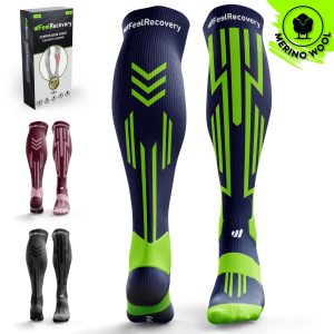
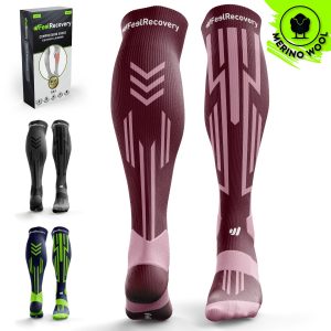
- Best products for foot fractures
- What are the causes and risk factors for foot fractures?
- First aid care for treating a foot fracture
- Most indicated treatments for a foot and ankle fracture
- Rehabilitation after a fracture of the foot bones
- Prevention methods to avoid broken bones in the feet
In general terms, a fracture is based on the loss of continuity of a bone due to its breakage. Among the most common types is the foot fracture, which is caused by numerous eventualities and triggers considerable discomfort and pain in the patient when trying to walk or bear weight on the limb.
Because it is a common injury that can occur in people of any age, it is worth knowing more about what it is. Therefore, we will highlight its main characteristics, how it is diagnosed, its classification, what causes it, how it should be treated and what ways exist to prevent it.
What is a fracture of the bones of the foot and how is it diagnosed?
A fracture of the bones of the foot is defined as a pathology that involves the breakage or fracture of a bone fragment linked to this limb and, as a result, causes a decrease in the range of movement of the foot.
It presents signs and symptoms such as:
- Pain (increases with walking)
- Swelling
- Bruising or discoloration of the skin
- Tenderness in the area
- Numbness of the foot or toes
- Crunching when moving the limb
- Visible deformity.
This injury can intervene in any area of the foot, either in the toes, the bones of the middle third of the foot, the two bones that are located under the toe or hallux and in the bones located in the upper part of the limb.
A foot fracture is commonly associated with:
- Phalanges: These refer to the bones located at the ends of the toes. They are long and allow the flexion, extension and opposition movements of the thumb. Normally, the fracture occurs in the big toe.
- Metatarsals: These are those bone fragments located in the middle of the foot that connect the ankle to the toes and help maintain balance when walking or standing. Generally, fractures in this bone are caused by overload, a sudden blow or a severe sprain.
- Sesamoids: These are two small, rounded or pea-shaped bones located at the base of the big toe joint. In most cases, these are broken by repetitive stress or overuse and thus, manifest immediate pain and swelling in the area.
- Hindfootbones: These refer to bones such as the scaphoid and talus. The scaphoid inserts into a tendon medially and the talus helps form the ankle joint as it fits into the tibia and fibula. This type of fracture can occur due to a number of factors.
To diagnose a foot fracture, as well as it is important to evaluate the patient's symptoms, it is also essential that the medical specialist performs a physical examination of the affected area. To do this, the focus is on palpating different parts of the foot to detect and control which points of pain exist and/or their precise location, which, at the same time, will allow the cause of the bone break to be determined.
An imaging test is essential to obtain more detail about the fracture internally, usually one or more of the following studies are suggested:
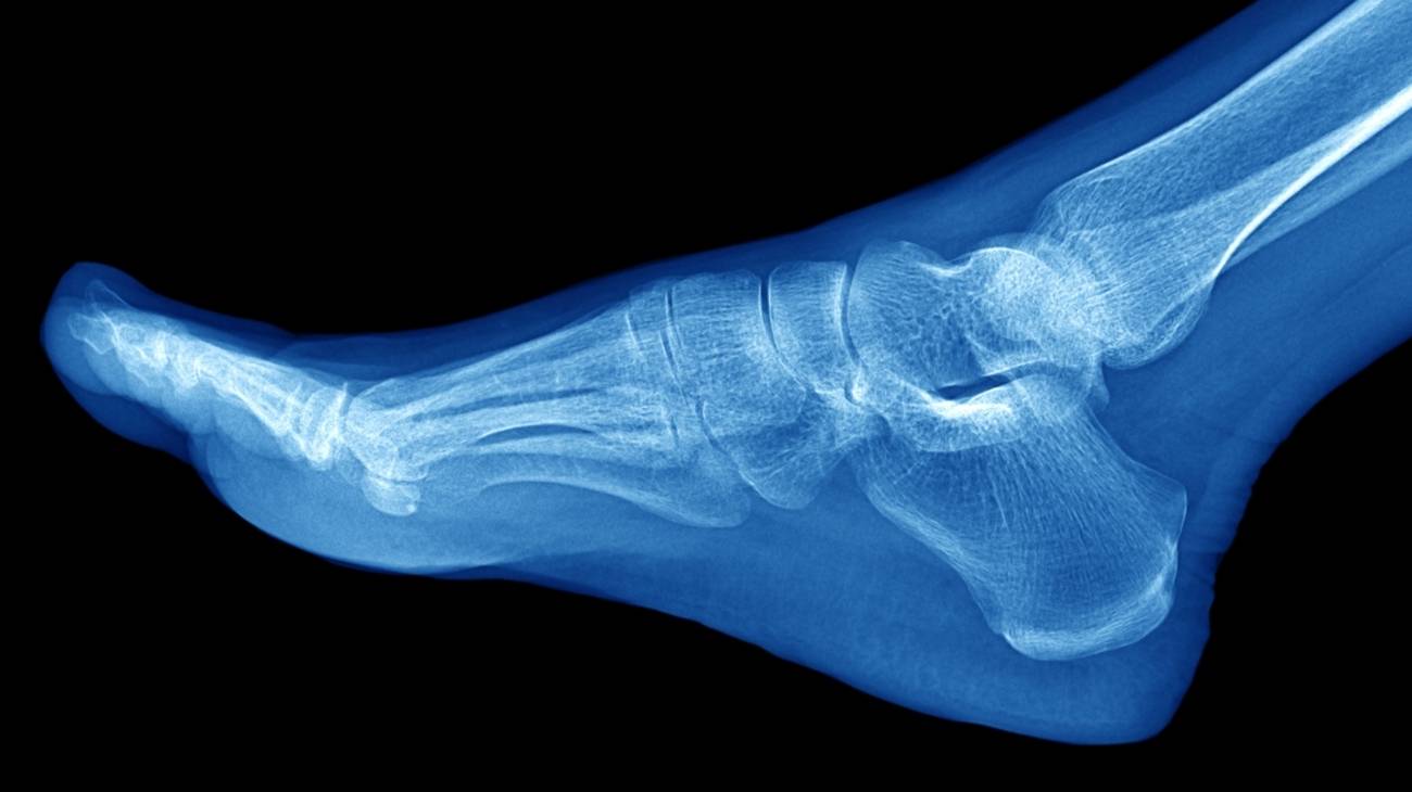
- X-ray: The X-ray test involves taking pictures from several different angles so that the images of the fractured bone do not overlap noticeably. In most cases, these types of foot injuries can be clearly visualised on X-rays, except for stress fractures which cannot be properly visualised until the break has begun to heal.
- Computed tomography: Similar to X-rays, this type of examination uses X-rays to produce images from various angles. Beyond this, however, it is distinguished by combining various techniques to create three-dimensional images of the internal structures of the body. As a result, it provides more detail about broken bones and even the surrounding soft tissues, allowing for more efficient treatment.
- Bone scan: To perform this imaging test, the technician begins by injecting a small amount of radioactive material into a vein. The radioactive material is then absorbed into the bones (especially the parts of the bone that are damaged). This makes it possible to visualise stress fractures with complete clarity in the resulting images, without the healing phase having been accessed.
- Magnetic resonance imaging: This uses radio waves together with a strong magnetic field to produce much more detailed imaging samples which, as well as exposing the condition of the bone fragments, also requires shots of the ligaments that help to support the foot and ankle in an interlocking fashion. As a result, it can identify fractures that are not visible on X-rays and even rule out any deterioration adjacent to the injured bone.
What are the types of foot bone fractures there are?

Since the structure of the foot is made up of different bones that make up a total of 30 joints supported by more than 100 bony ligaments, fractures in this limb are considered to be highly complex.
One of the most relevant classifications depends on the bone affected during impact:
- Talus fracture: This is a type of bone fracture in which the fracture line is in the talus bone located in the foot. In most cases, it is produced by a violent trauma that generates dorsal hyperflexion of the extremity and impacts the neck of the talus and the anterior border of the tibia. To heal it, a surgical reduction must be performed in order to install the bone fragments in their anatomical position.
- Calcaneal fracture: This is an injury that consists of the breakage of the homonymous bone located in the heel of the foot and is normally caused by a fall from a height in which the foot suffers a considerable contusion on contact with the ground. As they are complex tears, their evolution is very slow and sometimes leads to functional incapacity or permanent sequelae. Treatment may be based on immobilisation or surgery, depending on the severity of the case.
- Cuboid fracture: Although infrequent, they are vital in this classification. Basically, this fracture is triggered by direct trauma to the cuboid bone, which is the bone on the outer side of the midfoot. However, in certain cases, the cuboid is fractured as a result of a stress mechanism in military personnel or athletes (especially runners). This injury, in turn, is segmented into: simple fracture, intra-articular fracture and comminuted fracture.
- Scaphoid fracture: The scaphoid is a boat-shaped bone found in the midfoot or between the head of the talus and the three wedges, specifically. When such a bone break occurs, the symptoms experienced by the patient are usually mild and do not prevent continued regular activity of the limb. Generally speaking, this injury is caused by stress or overuse.
- Metatarsal fracture: The metatarsal bones are long and are responsible for connecting the ankle to the toes. They are also responsible for keeping a person's balance while standing or walking, so if a metatarsal fracture occurs, it is usually severe. Most often, broken metatarsal bones can be due to compression fractures or trauma fractures.
- Fracture of the phalanges of the foot: These bones allow flexion, extension and opposition movements of the thumb, as well as articulation with the metatarsals. Once a fracture develops in the foot, they can break because they are small and fragile. Thus, they cause significant pain and swelling that can take up to 4 to 6 weeks to completely disappear.
- Chopart's dislocation fracture: The Chopart's joint is primarily characterised by the taloscaphoid and calcaneocuboid joints. Thus, if certain injuries to these joints occur, a rupture of the ligaments is likely to occur, resulting in a fracture of the scaphoid or cuboid in the impacted foot. However, these are very rare breaks that occur in very few cases.
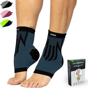
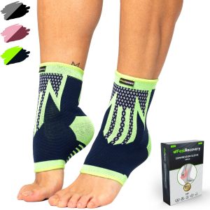
Best products for foot fractures
Bestseller
-

2 Ankle Compression Sleeve (Black/Gray)
£17,50 -
2 Ankle Compression Sleeve (Green/Navy)
£17,50 -
2 Ankle Compression Sleeve (Pink/Bordeaux)
£17,50 -
Acupressure Mat and Pillow (Black/Gray)
£44,95 -
Acupressure Mat and Pillow (Green/Navy)
£44,95 -
Acupressure Mat and Pillow (Pink/Bordeaux)
£44,95 -
Acupressure Pillow (Black/Gray)
£21,52 -
Acupressure Pillow (Green/Navy)
£21,52 -
Acupressure Pillow (Pink/Bordeaux)
£21,52 -
Electric Foot Massager
£43,07 -

Foot Massage Roller for Plantar Fasciitis (Black)
£17,50 -
Foot Massage Roller for Plantar Fasciitis (Green)
£17,50 -
Foot Massage Roller for Plantar Fasciitis (Pink)
£17,50 -
High Density Foam Roller for Muscle (Black/Gray)
£24,95 -
High Density Foam Roller for Muscle (Green/Navy)
£24,95 -
High Density Foam Roller for Muscle (Pink/Bordeaux)
£24,95 -
Ice Massage Roller Ball (Black)
£34,95 -
Ice Massage Roller Ball (Green)
£34,95 -
Ice Massage Roller Ball (Pink)
£34,95 -
Ice Pack for Foot - Cold Therapy Socks (Black)
£21,95 -
Ice Pack for Foot - Cold Therapy Socks (Green)
£21,95 -
Ice Pack for Foot - Cold Therapy Socks (Pink)
£21,95 -
Microwavable Heated Slippers (Hearts)
£21,50 -
Microwavable Heated Slippers (Oxford)
£21,50 -
Microwavable Heated Slippers (Sport)
£21,50 -
Microwaveable Wheat Bag for Pain Relief (Hearts)
£17,50 -
Microwaveable Wheat Bag for Pain Relief (Oxford)
£17,50 -
Microwaveable Wheat Bag for Pain Relief (Sport)
£17,50 -
Pack 2 in 1: Foam Roller High + Soft Density (Black/Gray)
£24,95 -
Pack 2 in 1: Foam Roller High + Soft Density (Green/Navy)
£24,95 -
Pack 2 in 1: Foam Roller High + Soft Density (Pink/Bordeaux)
£24,95 -
Pack 3 Spiky Massage Balls
£17,50 -
Pack Lacrosse Massage Balls & Peanut Roller
£24,95 -
Reusable Gel Ice Packs for Ankle & Foot with Compression Band
£17,50 -
Reusable Gel Ice Packs with Compression Band for Sports Injury
£17,50 -
Soft Density Foam Roller for Recovery (Black)
£24,95 -
Soft Density Foam Roller for Recovery (Green)
£24,95 -
Soft Density Foam Roller for Recovery (Pink)
£24,95 -
Sport Compression Socks (1 Pair) (Black/Gray)
£17,50 -
Sport Compression Socks (1 Pair) (Green/Navy)
£17,50 -
Sport Compression Socks (1 Pair) (Pink/Bordeaux)
£17,50 -
Vibrating Foam Roller
£25,83 -
Vibrating Massager Ball
£25,83


What are the causes and risk factors for foot fractures?
The frequency with which foot fractures occur in emergency rooms is due to the fact that this type of injury is caused by numerous eventualities. In other words, both the causes and risk factors vary and are essential to analyse each patient's case, provide a concise diagnosis and formulate a good treatment.
The most common factors that increase the risk of suffering a rupture of this type are:
- Falls are the main eventualities that cause a foot fracture. For example, a stumble that causes you to fall to the ground abruptly, or landing on your feet after jumping from any height, affects the bones of the limb directly.
- People who suffer blows or trauma to the foot are also prone to severe injuries to the area, especially bone fractures.
- The impact of a heavy object or dropping something hard on the foot causes a bone in the lower limb to break. This is one of the most common causes.
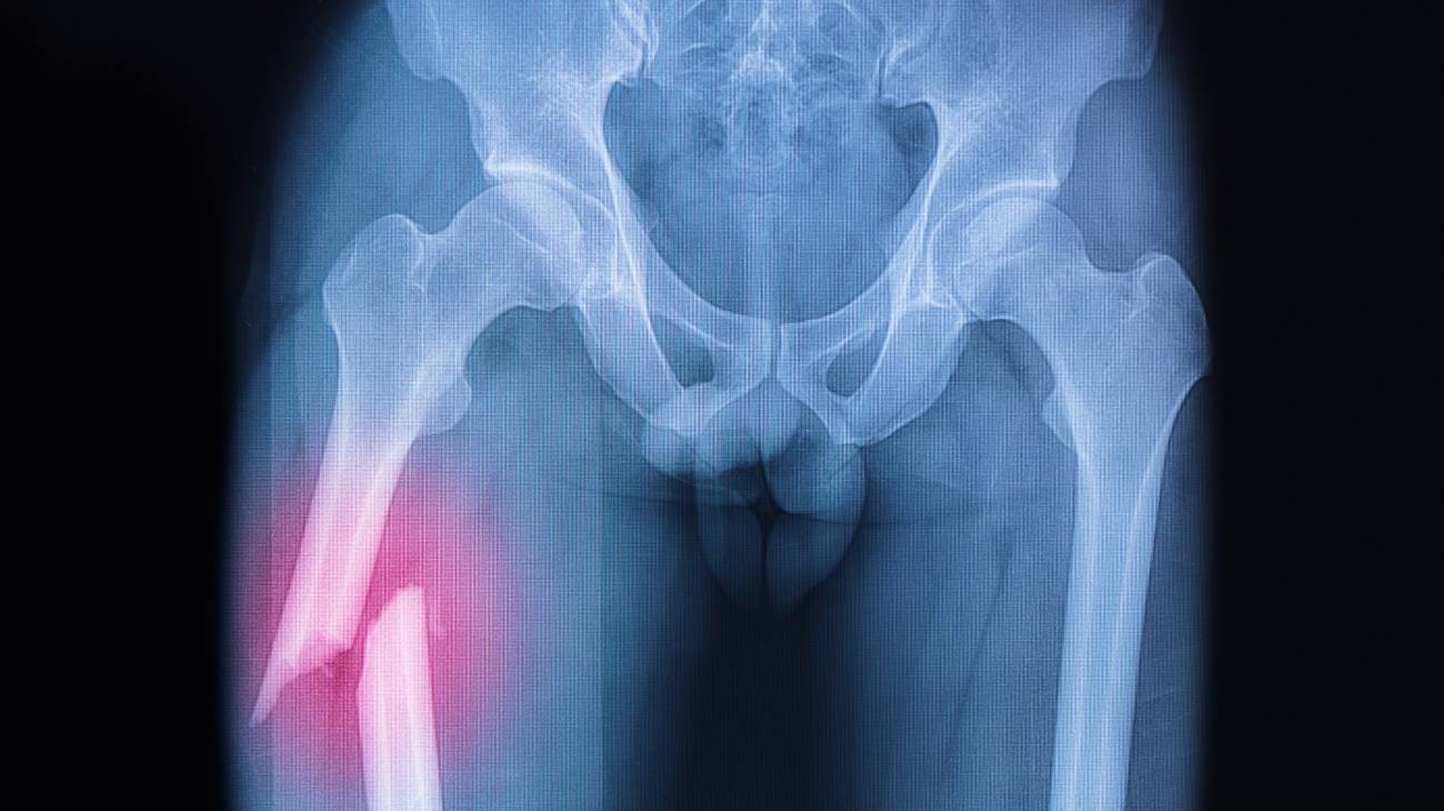
- Traffic accidents (by car, motorbike or bicycle) increase the risk of having to endure a broken bone in the foot. Since, collisions often develop high-energy fractures and crush injuries.
- Slips that an individual slips while running or walking and steps wrongly on the ground or places the foot in an incorrect posture cause twisting tears that usually break bones.
- Overuse: Which, due to repetitive force or formidable wear and tear, tends to lead to stress fractures in the weight-bearing bony fragments of the feet.
- Increasing the level of activity while exercising suddenly, in terms of duration, intensity and/or frequency, can trigger a stress fracture in the foot area. As well as the use of contraindicated equipment and inappropriate movements during the practice of any sport or skill.
- Osteoporosis or congenital bone pathologies tend to produce fractures in the feet and in any region of the body easily.
- Athletes involved in running, gymnastics, tennis and basketball, i.e. sports with high impact or repetitive movements, are considered a population at prominent risk for foot bone fractures.
- Advancing age is identified as another cause of this type of injury. As the bones become weaker and more fragile over the years, they are prone to break easily.
- People who have to work in certain work environments that involve a risk of falling from a certain height or suffering an impact caused by heavy objects on the feet (a construction site, for example), can experience a fracture in this part of the body at any time.
- Bad habits (smoking and drinking alcohol) increase the chances of having a bone fracture in the foot. Smoking stimulates the development of osteoporosis in individuals and alcohol causes bone loss, as well as disorientation and thus leads to falls or traffic accidents.
- Bones can also be weakened by a deficiency of certain nutritional supplements in the body. Consequently, another reason for a foot fracture is due to poor nutrition.
First aid care for treating a foot fracture
When a foot fracture is exposed, the patient is unable to walk or move the limb. Consequently, the first step in first aid is to call for trained medical help to assist the patient correctly.
However, during the time it takes to receive clinical care, it is essential that the injured person and those trying to help them take into account certain steps or precautions to prevent the fracture from worsening and putting the patient's health at risk for the long term.
It is appropriate to implement the following primary care measures:
- Immobilising the limb: Moving broken bones increases pain and inflammation, as well as causing damage to the tissues around the injury. Therefore, it is vital to immobilise the foot quickly to protect it from further damage and when doing so, you can place a garment or folded blanket between the lower limbs (feet together and wrapped) to prevent twisting movements of the broken bone.
- It is not appropriate to touch or try to align the broken bone fragment: When immobilising the fractured foot, you should avoid aligning the broken bone, pushing it inwards and touching it, i.e. it should be left as it is after the impact. Otherwise, it is possible that it will attract a bone infection and increase its level of severity, especially if it is an open or exposed fracture (the bone protrudes through the skin).
- Monitor the affected person's vital signs: As far as possible, it is essential to carry out a primary assessment that consists of monitoring the patient's vital signs or checking how the indicators that provide information about their state of health are behaving. In this regard, it is advisable to take their pulse, take their temperature, measure their respiratory rate, blood pressure and blood pressure, as well as check that they are conscious. It is also valuable to prevent them from falling asleep or closing their eyes.
- Provide thermal protection: It is also important to prevent the casualty from losing too much heat. Therefore, to prevent their temperature from rising noticeably, you can cover or tuck the patient in a blanket over the entire length of their body.
- Over-the-counter medication is not recommended: It is not ideal for a recently fractured patient to self-medicate with over-the-counter drugs to reduce the pain in the affected area or to eliminate the swelling that can be observed. This tends to trigger adverse health effects and can worsen the patient's condition with vomiting, diarrhoea, fainting, drowsiness and even cardiac arrest. It is also important to avoid consuming fluids or food while in care.
- Apply cold compresses to the fractured region: Cold has natural analgesic, anti-inflammatory and sedative properties. It is therefore appropriate to place cold compresses on the affected foot so that the patient can obtain some relief from the discomfort he or she feels. When doing so, it is not appropriate to put ice directly on the skin as this will almost always cause burns.
- Control the person's nervousness: Due to the impact suffered, the injured individual is likely to go into a state of shock and nervousness that can aggravate the condition of the fractured bone and increase the pain exponentially. For this reason, it is recommended to help the patient to relax or feel calm in order to favour their stability while they can obtain the requested medical help.
The action of the first aid team may follow the following pattern:
- Immobilise the foot with the appropriate elements.
- Maintain stability of vital signs and organ function.
- Administer a strong analgesic to help relieve pain.
- Control bleeding, if there is an open fracture with wound.
- Order an X-ray of the foot where the fracture is suspected.
Most indicated treatments for a foot and ankle fracture
The treatment to improve a foot and ankle fracture will depend on the bone that was injured and the type of fracture the patient developed. Thus, depending on the diagnosis and based on the level of severity of the bone break, medical specialists will be able to determine which procedure the patient should follow to improve their foot fracture.
To prevent harmful side effects of drug treatment, it is valuable to take into account certain details about each individual when writing a prescription, such as: medical history, health status, drug tolerance, allergies, age, etc. Otherwise, it is likely that the patient will develop other pathologies that may impair his or her evolution and quality of life (dizziness, fainting, vomiting, diarrhoea, drowsiness, addiction, cardiac arrest, etc.).
The foot reduction and immobilisation procedures consist of:
- Reduction: If the fracture is displaced (or the two ends are not aligned), the specialist in the area will have to manipulate the bony portions back into their correct position. Depending on the swelling observed and the patient's tolerance to pain, a muscle relaxant, sedative or general anaesthetic may be needed to ensure that the treatment is not painful.
- Immobilisation: In order to heal, it is vital that the fractured bone be subjected to a regime of immobilisation for a certain period of time. For this, it is indicated to place a cast or splint along the foot and ankle in order to limit movement in the whole area. In some cases, doctors may even suggest only fitting a special stiff-soled boot or shoe that leaves the toes free to prevent a more serious fracture. Through immobilisation, the broken bone ends will be able to knit back together.
It is essential to avoid supporting the limb for long periods of time, as well as to avoid any weight bearing because this could intervene detrimentally in the healing progress. The time the patient must wait before being able to move the foot and walk normally again will depend on the type of injury and, depending on this, tends to last for weeks or even months.
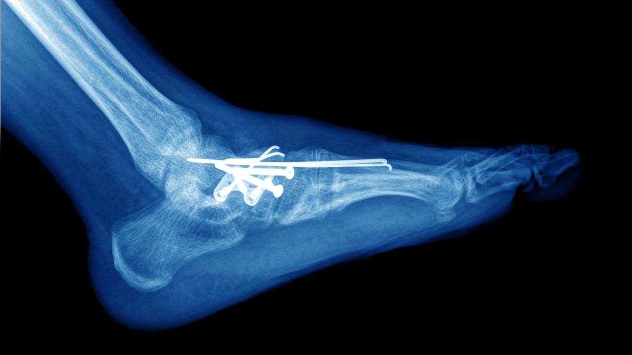
If the affected foot fails to recover with the above-mentioned treatments, the fracture suffers a complication and is truly severe, surgical treatment is considered mandatory. In such cases, the surgeon will opt to use metal plates or screws to help preserve the exact anatomical position of the broken bones during the healing time.
Rehabilitation after a fracture of the foot bones

After treatment (conservative or surgical) has been completed, the next step is to begin the rehabilitation process to improve the movement and flexibility of the injured foot, as well as to strengthen all of its muscles. When the doctor determines that the bone has welded in place, he or she will indicate that the patient should undergo a physiotherapy plan that will make the regeneration of the limb much more effective.
The physiotherapist will establish a plan with personalised exercises to help relieve pain, optimise range of motion, stimulate elasticity and tone muscles.
- Phalanges and metatarsals can heal in 3 to 6 weeks.
- The tarsal bones take longer, 6 to 10 weeks.
The most common procedures to be followed to accelerate rehabilitation after a fracture of the bones of the foot are:
- Mechanotherapy: This is a type of therapeutic technique that involves passive, active or active-assisted mobilisation, depending on the condition of the fractured area. Apart from this, it promotes strengthening exercises, resistance exercises, balance exercises and proprioception practices.
- Electrotherapy: This is a post-treatment that uses analgesic and anti-inflammatory currents to eliminate the main signs and symptoms of a bone fracture of this type. It also applies ultrasound and laser to strengthen the injured region.
- Hydrotherapy: Consists of the use of water as a therapeutic agent and to rehabilitate a foot after suffering a fracture, it normally makes use of a tank and a whirlpool bath suitable for triggering good results.
- Cryotherapy and thermotherapy: These are physiotherapies that use the properties of cold and heat, respectively, to attenuate the discomfort caused by a fracture. Normally, they are applied with cold compresses and hot wet compresses directly on the affected area for a certain period of time that is defined by the physiotherapist.
- Gait re-education: This is an essential procedure that stands out for the set of actions designed to enable a patient to walk again efficiently and safely, with excellent mobility, strength, coordination, balance and proprioception. These practices prevent the condition from persisting with chronic pain or swelling in other segments of the foot, leg, knee and even hip.
Prevention methods to avoid broken bones in the feet
We advise you to implement the following suggestions in order to preserve the health of your bones in your feet as well as in any other part of your body:
- Engage in physical activity steadily and gradually: While it is true, sport has the ability to strengthen muscles, stimulate joint health and even increase the amount of bone, so it is advisable to practice physical activities to minimise the risks of suffering a bone fracture. Bearing in mind that, just as it is essential to do it day after day, it is considered vital to do it gradually to prevent overload injuries due to exorbitant training.
- Alternating sporting activities: Another way to avoid stress fractures is to alternate training so that the bones of the lower limbs (especially the feet) do not break suddenly. A clear example of this is based on walking, running, swimming and cycling in turn.
- Wearing the right shoes: In order to perform certain skills as well as to avoid falling at any time, it is essential to wear shoes that are suitable for your feet, i.e. that fit perfectly. They should also be suitable for each type of sport and even have a non-slip sole. In addition, it is advisable to replace sports shoes periodically, especially if they show irregular wear.
- Having a healthy and balanced diet: It is essential to follow a healthy and balanced diet that helps the body to absorb all the nutrients and substances it needs to keep it functioning properly. In order to support the stimulation of bone strength and avoid bone weakness, it is suggested to eat foods with a high content of calcium, vitamin D and collagen. Likewise, to avoid a deficit of these substances, it is possible to take nutritional supplements that provide these with their properties.
- Avoid habits harmful to health: Another useful formula for obstructing the manifestation of bone fractures consists of preventing harmful habits, such as: sedentary lifestyles, eating disorders, smoking tobacco and drinking alcohol. Since, this stimulates the weakening of bones, favours the development of osteoporosis, causes bone loss and facilitates falls or sudden eventualities.
- Implement safety tips for outdoor activities: To prevent falls, trips and/or subjective accidents, it is appropriate to carry out the following: Avoid walking in dark or poorly lit places, keep your hands free, walk with great caution in buildings with marble or highly polished tile floors, use a cane if necessary and drive respecting traffic signs.
- Keep the floor of your home clear: If you want to minimise the risk of a fall that could break your foot, we also suggest that you keep the floor of your home completely conditioned so that you can walk without tripping. To do this, it should be free of clutter, not cleaned with slippery wax, free of shifting rugs or loose wires, and keep furniture or decorative objects in the right place.
- Leaning on handrails when going up/down stairs: Many people often roll down stairs because they do not lean on handrails and as a result, suffer bone fractures involving the bones of the foot. If you have stairs in your home, be sure to install handrails on both sides, tape bright tape to differentiate the steps easily and install light switches to keep the area well lit.
- Clean up debris in the kitchen area of your home: To prevent falls or household accidents, it is also advisable to clean up liquid or food spills once they occur in the kitchen (or anywhere else in the house). It is effective to place non-slip mats near the cooker and sink.
References
- Bica, D., Sprouse, R. A., & Armen, J. (2016). Diagnosis and management of common foot fractures. American family physician, 93(3), 183-191. https://pubmed.ncbi.nlm.nih.gov/26926612/
- Ribbans, W. J., Natarajan, R., & Alavala, S. (2005). Pediatric foot fractures. Clinical Orthopaedics and Related Research®, 432, 107-115. https://journals.lww.com/clinorthop/Fulltext/2005/03000/Pediatric_Foot_Fractures.14.aspx
- Seeley, D. G., Kelsey, J., Jergas, M., & Nevitt, M. C. (1996). Predictors of ankle and foot fractures in older women. Journal of Bone and Mineral Research, 11(9), 1347-1355. https://asbmr.onlinelibrary.wiley.com/doi/abs/10.1002/jbmr.5650110920
- Keegan, T. H., Kelsey, J. L., Sidney, S., & Quesenberry Jr, C. P. (2002). Foot problems as risk factors of fractures. American Journal of Epidemiology, 155(10), 926-931. https://academic.oup.com/aje/article/155/10/926/144154
- Hasselman, C. T., Vogt, M. T., Stone, K. L., Cauley, J. A., & Conti, S. F. (2003). Foot and ankle fractures in elderly white women: incidence and risk factors. JBJS, 85(5), 820-824.
- Klotzbuecher, C. M., Ross, P. D., Landsman, P. B., Abbott III, T. A., & Berger, M. (2000). Patients with prior fractures have an increased risk of future fractures: a summary of the literature and statistical synthesis. Journal of bone and mineral research, 15(4), 721-739. https://journals.lww.com/jbjsjournal/Abstract/2003/05000/Foot_and_Ankle_Fractures_in_Elderly_White_Women___.8.aspx https://asbmr.onlinelibrary.wiley.com/doi/full/10.1359/jbmr.2000.15.4.721
- Court-Brown, C. M., & Caesar, B. (2006). Epidemiology of adult fractures: a review. Injury, 37(8), 691-697. https://www.sciencedirect.com/science/article/abs/pii/S0020138306003238
- Singer, B. R., McLauchlan, G. J., Robinson, C. M., & Christie, J. (1998). Epidemiology of fractures in 15 000 adults: the influence of age and gender. The Journal of bone and joint surgery. British volume, 80(2), 243-248. https://online.boneandjoint.org.uk/doi/abs/10.1302/0301-620X.80B2.0800243
- Bischoff-Ferrari, H. A., Willett, W. C., Wong, J. B., Giovannucci, E., Dietrich, T., & Dawson-Hughes, B. (2005). Fracture prevention with vitamin D supplementation: a meta-analysis of randomized controlled trials. Jama, 293(18), 2257-2264. https://jamanetwork.com/journals/jama/article-abstract/200871
- Patel, D. S., Roth, M., & Kapil, N. (2011). Stress fractures: diagnosis, treatment, and prevention. American family physician, 83(1), 39-46. https://www.aafp.org/pubs/afp/issues/2011/0101/p39.html