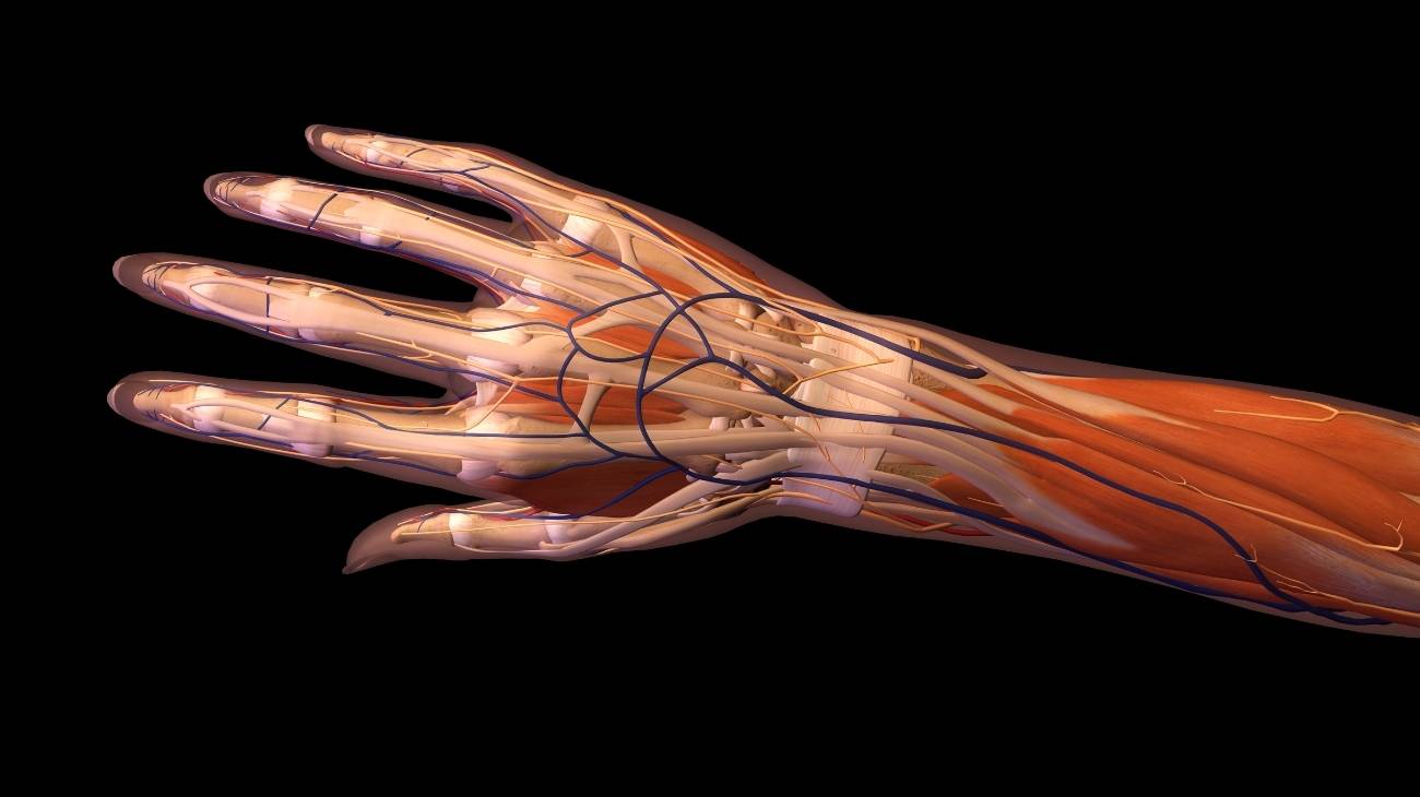
The anatomy of the wrist, hand and fingers is very complex, because it involves 27 bones and a large number of muscles and ligaments that are used in biomechanical movements. This joint part of the human body is prone to all types of injury, from fractures, tendonitis and even bursitis.
If you want to know more about the treatments that exist to put wrist conditions into remission, it is important that you continue reading. We will explain in detail all the complementary and non-invasive therapies you can choose from. In addition, you will find the best products to help you fight the pain.
Parts and anatomy of the hand
The anatomy of the hand is composed of the following parts:
Bones and joints
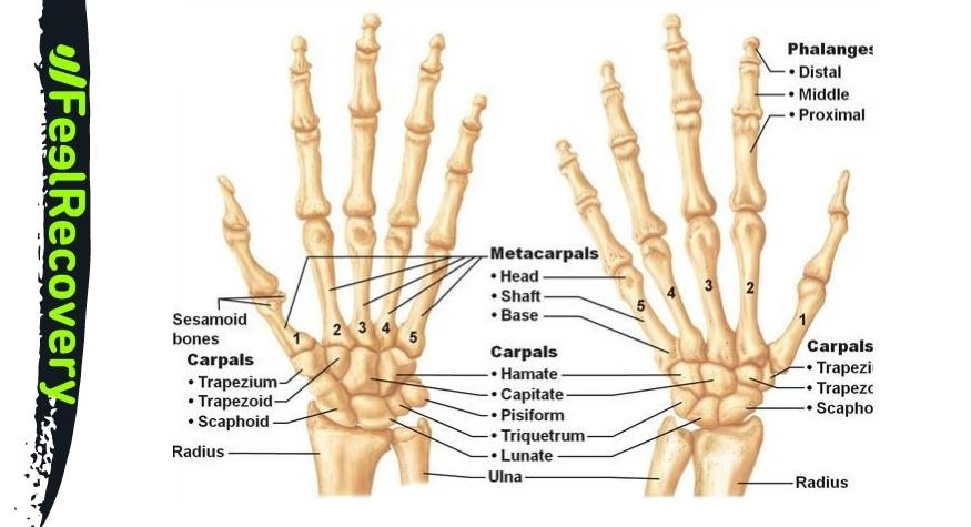
The bones that make up the wrist can be divided into two groups according to their location in relation to the radius and ulna.
If the proximal distance of this bony structure from the forearm is taken into account, the bones of the wrist are
- Scaphoid: 3 of its 6 faces are articular, so it has contact with the radius bones of the forearm, the lunate, the trapezium, the trapezoid, the trapezium and the great bone. It is the bone most likely to fracture.
- Semilunate: Its name derives from the shape of this bone, which is compact, cuboid and spongy. Its facets are related to the radius, large, hooked, scaphoid and pyramidal. Two of these facets are not articular.
- Pyramidal: Like the previous bone, this bone tissue has its name due to its shape. It has 6 facets, 3 of which are articular. It connects with the pisiform, lunate and hooked.
- Pisiform: This bone is located on the outside of the wrist and may form the group with distal location, but it is considered within this sector. It is next to the hook and pyramidal bones. It is a strategic bone because it contains the little finger adductor muscle and the anterior ulnar muscle. Its four facets give rise to the ulnar nerve and the artery of the same name.
Considering the distal distance, the bony structure of the wrist consists of
- Trapezius: Articulates with the first metacarpal of the thumb. It also connects with the scaphoid and trapezium. It is part of the important trapezometacarpal joint.
- Trapezoid: Articulates with the second metacarpal of the index finger. Of its 6 facets, 4 are articular because it connects with the trapezium, great and scaphoid bones.
- Great: This bone articulates with the second, third and fourth metacarpal bones of the middle or middle finger. Four facets are articular and it is related to the scaphoid, lunate, trapezium and hooked bone.
- Ganchus: It is responsible for the articulation of the fourth and fifth metacarpals of the ring and little fingers. It is related to the large bone, the pisiform, the pyramidal and the lunate.
In terms of the bones that make up the hand, the following can be distinguished from a proximal to distal course:
- Metacarpals: Also called metacarpals, they are five bones and form the palm of the hand. They are counted from the thumb to the little finger. They are responsible for producing the articulation between the palm of the hand and the wrist. They are covered with tendons that restrict the movements of the fingers.
- Proximal phalanx: Corresponds to one for each finger. They are related to each of the metacarpals, forming joints between the metacarpals and the middle phalanges.
- Middle phalange: In this case the number of bones is less than the other phalanges, because the thumb does not have this bone tissue. Its action is to produce movements between the proximal phalanx and the distal phalanx of each finger.
- Distal phalanx: This bone is found one for each finger, forming the last link of the digital bones. This bone may also be referred to as the phalanx or nail phalanx. It articulates with the middle phalanx and in the case of the thumb with the proximal phalanx.
- External sesamoid: A small bone that lies between different joints in the hands, its job is similar to that of a pulley. This means that it reduces the tension on the ligaments and other muscles when certain movements are performed.
One of the characteristics that distinguishes humans from other species is their ability to articulate the 27 bones of their hands. We will show you below what these articular bodies are and what they are called.
These joints are found in the wrist:
- Radio ulnar: This joint allows movement of the ulna and radius of the forearm, which helps the radiocarpal joint.
- Radiocarpal: This body allows movement between the radius and the scaphoid and the radius with the lunate. It is common to find the name of each of these joints called radioscaphoid and radiosemilunar.
- Intercarpal: The movement of the lunate bone, with the piriformis, pisiform, hooked bone and large bone are produced by the action of this joint.
- Carpometacarpal: These joints are found at the junction of the hook bone, the large bone, the trapezium and the trapezium with the metacarpals of the hand. It is possible to distinguish each of these joints by linking the names of the bones they join.
The joints of the hand are:
- Carpometacarpal: This is the joint that makes it possible to move the carpal bones with each of the metacarpals of the palm of the hand.
- Metacarpophalangeal: The joint between the metacarpals and the proximal phalanges can move thanks to this joint.
- Proximal interphalangeal: This joint can be found at the junction of the proximal and middle phalanges.
- Distal interphalangeal joint: This joint allows the movement of the middle phalanges with the distal phalanges of the fingers.
- Proximal-distal interphalangeal joint: Unlike the previous articular bodies, this joint is present in each hand, as it is responsible for moving the proximal phalanx with the distal phalanx of the thumb.
Muscles
The muscles of the hand and wrist are
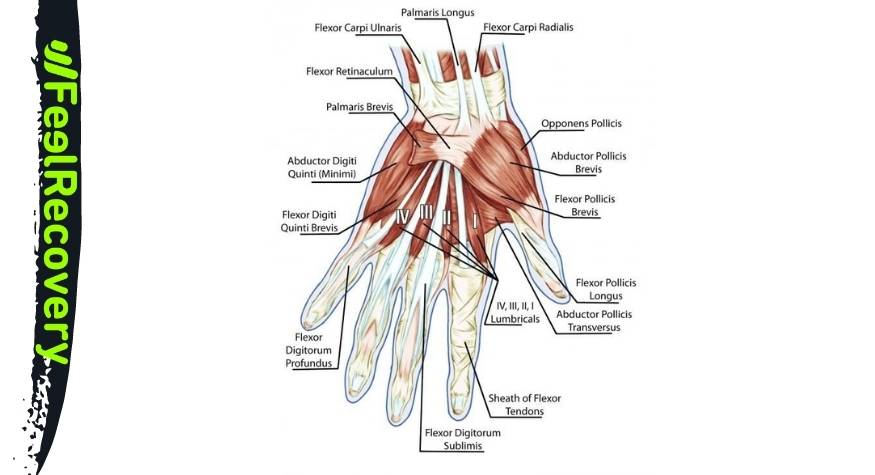
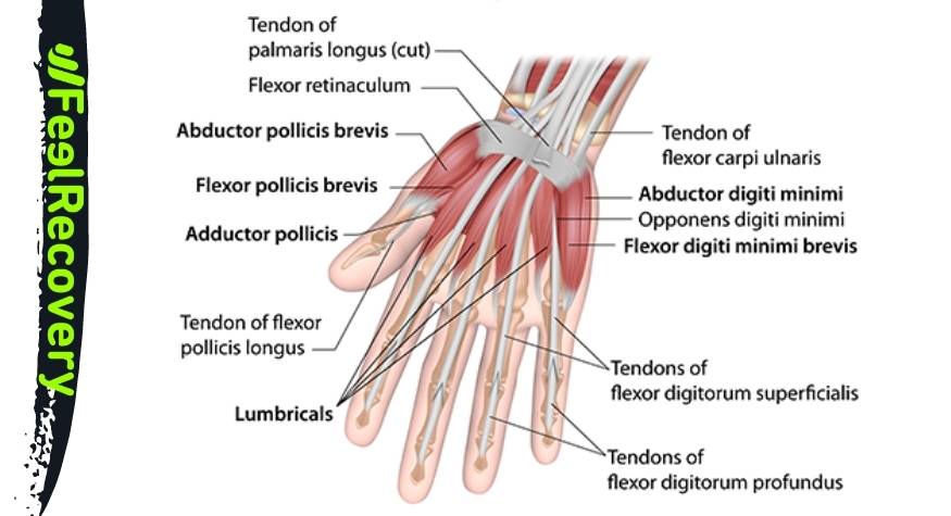
- Opponent of the thumb: It arises from the tubercle and tensors of the trapezius and the annular ligament of the carpus. It inserts on the first metacarpal. Its action is to rotate the thumb to touch the palm of the hand and the other fingers.
- Abductor pollicis brevis: It travels from the scaphoid and the annular ligament to the first phalanx of the thumb. Its job is to pull the thumb away from the palm of the hand.
- Flexor pollicis brevis: This muscle arises from the large bone, the flexor retinaculum and the trapezius tubercle. It attaches to the sesamoid and the first phalanx. Its downward movement consists of flexion of the thumb.
- Adductor pollicis: It arises from the first row of the carpals, on the scaphoid. It inserts on the opponent of the thumb reaching the external tubercle of its base. The action of this muscle is to control adduction movements.
- Little finger abductor: It arises from the pisiform bone and inserts on the fifth proximal phalanx of the little finger. Its function is to balance the movements of the little finger.
- Little finger flexor digitorum brevis: This muscle can be found from the hooked process to the ulnar border of the first phalanx of the little finger. Its action is to maintain balance in the flexion of this finger.
- Opponent of the little finger: It arises from the hook bone and inserts into the diaphysis of the fifth metacarpal. Its function is to rotate the little finger when it faces the thumb.
- Lumbrical: It is a set of 4 muscles located in the dorsal part of the hand. They arise from each of the deep flexors of the fingers to flex and extend the first phalanx.
- Palmar interosseous: This is also a set of muscles, but in this case there are three. They originate from the metacarpals to the proximal phalanges of the index, ring and little fingers.
- Dorsal interosseous: This muscle is found in each of the fingers from the metacarpals to the first phalanx. It regulates abductor and flexor movements of the hand.
- Ulnar flexor carpi ulnaris: This is a muscle of the forearm that attaches to the pisiform, hook and fifth metacarpal. Its action is to flex and control wrist movements.
- Flexor superficialis: The action of these muscles is to flex the forearm and fingers. The path of this tissue originates in the coronoid process of the ulna and in the medial epicondyle of the humerus up to the ligament of the middle phalanges.
- Deep flexor: These muscles have their origin in the interosseous ligament, ulna and antebrachial aponeurosis. They insert on the third phalanx of the fingers, except for the thumb.
- Palmar brevis: It is an internal muscle and small in size. It originates in the palmar aponeurosis and inserts into the deep part of the skin layer, called the dermis.
- Palmaris major: It originates in the epitrochlea of the elbow and inserts into the second metacarpal of the hand. Flexion of the wrist is related to the action of this muscle.
Ligaments
The ligaments found in the wrist and hands from the palmar side are:
- Tissues that join the radius to the carpal and metacarpal bones: radioscaphocapitate, radioscaphosemilunate, triquetral radiolunate, ulnotriquetral, ulnolunate and triquetocapitate. Their function is to give stability to the wrist and to limit some movements.
- Ligaments that hold the metacarpal and carpal bones to the ulna of the forearm: ulnar piriformis and ulnar lunate.
Considering the dorsal side of the hand and wrist, it is possible to find the following ligaments
- Dorsal intercarpal: This is responsible for joining the lunate bone to the scaphoid.
- Radiopyramidal: This ligament joins the radius bone with the pyramidal bone to limit and stabilise the wrist.
- Radiosemilunar: The lunate bone is attached to the forearm by this ligament.
- Radioscaphoid: The scaphoid and radius are joined by this tissue.
- Radial collateral: This is a ligament that arises in the elbow area and works with the thumb joint from the radius.
- Ulnar collateral: The thumb also receives the insertion of this ligament which arises from the ulnar bone of the forearm. Its job is to flex and extend the right finger.
Best products for hand, wrist and finger pain relief
Bestseller
-
Acupressure Mat and Pillow (Black/Gray)
£44,95 -
Acupressure Mat and Pillow (Green/Navy)
£44,95 -
Acupressure Mat and Pillow (Pink/Bordeaux)
£44,95 -
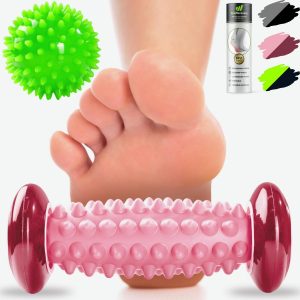
Foot Massage Roller for Plantar Fasciitis (Black)
£17,50 -
Foot Massage Roller for Plantar Fasciitis (Green)
£17,50 -
Foot Massage Roller for Plantar Fasciitis (Pink)
£17,50 -
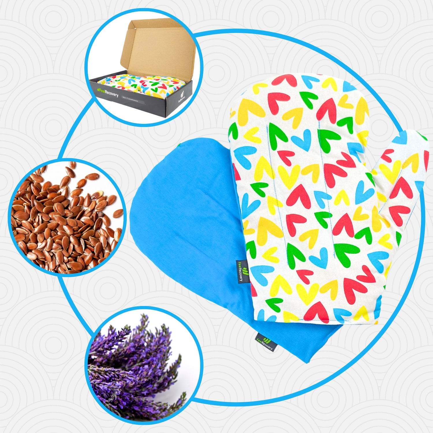
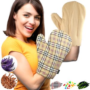
Microwave Arthritis Gloves (2 Mittens) (Hearts)
£25,50 -
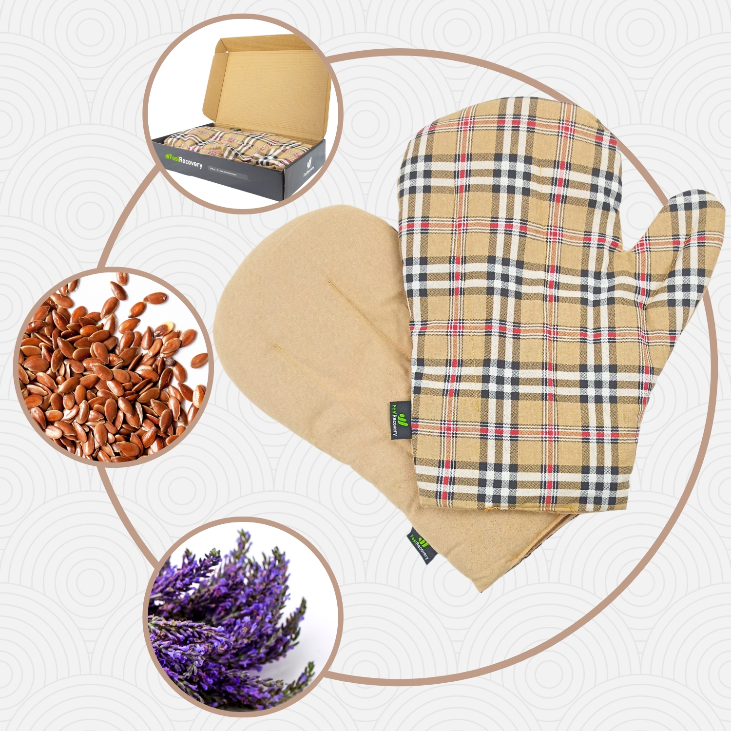
Microwave Arthritis Gloves (2 Mittens) (Oxford)
£25,50 -
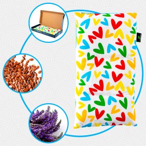
Microwaveable Wheat Bag for Pain Relief (Hearts)
£17,50 -
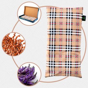
Microwaveable Wheat Bag for Pain Relief (Oxford)
£17,50 -
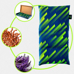
Microwaveable Wheat Bag for Pain Relief (Sport)
£17,50 -
Wrist Brace (Black/Gray)
£17,50 -
Wrist Brace (Green/Navy)
£17,50 -
Wrist Brace (Pink/Bordeaux)
£17,50
Biomechanics of the hand, fingers and wrist
The movements of the wrist and hand according to the biomechanics are as follows
- Flexion: This action occurs when the palm of the hand is brought towards the front of the forearm. This is achieved thanks to the tension produced by the ligaments located in the pyramidal, trapezium and trapezoid bones to move the scaphoid bone. The range of motion can be up to 90°.
- Extension: This is the opposite of flexion. It consists of placing the hand, in its dorsal area, as close as possible to the upper part of the forearm. This movement can be executed with an amplitude of up to 85°. The radius, lunate and scaphoid are the bones that act with the ligaments for this task.
- Abduction: This consists of placing the palm of the hand towards the ground and moving it parallel to the ulna and radius towards the trunk. This is achieved between 15 and 25° of amplitude thanks to the joints that move the carpal bones, the scaphoid and the lunate.
- Adduction: This action tries to position the hand in the same way as in abduction, but turn it to the opposite side. In other words, moving towards the outside of the trunk of the body.
The biomechanics of the body also allows movements in the fingers, which we detail below
- Abduction: When the fingers of the hand are separated, achieving the maximum opening, this arc is known as abduction.
- Adduction: This involves bringing the four fingers together while keeping the thumb separated from the palm of the hand.
- Extension: If the hand is placed in the position of adoption and the thumb is moved towards the palm, the extension movement is produced.
- Flexion: Moving the five fingers towards the palm allows the flexor muscles of the hand to perform this biomechanical action.
- Palmar: This movement allows the fingerprints of the index finger to touch with that of the thumb.
- Lateral: This biomechanical action places the thumb next to the phalanx of the index finger.
- Interphalangeal thumb flexion: It is possible to move the thumb between the two phalanges so as to form a 90° angle. Extension will be the opposite movement.
- Metacarpophalangeal flexion of each finger: Moving each of the fingers at 90 degrees in relation to the phalanx with the metacarpals causes this movement. Metacarpophalangeal extension is the opposite action to this biomechanical task.
- Proximal and middle interphalangeal flexion of each finger: When each finger is moved between the proximal and middle phalanges it is known as flexion (or extension, in the opposite case) of the fingers.
- Distal interphalangeal flexion: To perform this movement it is necessary to bend the middle and distal phalanges while keeping the proximal phalanx straight in relation to the metacarpals. This movement can be performed on all fingers except the thumb.
- Metacarpophalangeal adduction: This movement consists of moving each of the fingers towards the outside of the hand between the proximal phalanges and the metacarpals. Returning to the initial position is known as metacarpophalangeal abduction.
- Cylindrical grip: This consists of keeping the four fingers curved and the thumb underneath, as is done when grasping a cylindrical object.
- Hook: Unlike the cylindrical grip, the thumb is moved away from its lower position. That is to say, it consists of picking up all the fingers and leaving the thumb raised.
- Tip grip: This movement differs from the palmar grip in that it is not the fingerprints that are touched, but the ends of the fingernails.
- Spherical: Very similar to the cylindrical grip, but in this case the palm of the hand is facing upwards.
Most common hand and wrist injuries
See below for the most common types of hand, finger and wrist injuries.
Types of Hand, Finger and Wrist Injuries
Find out about injuries caused by different diseases:
- Arthrosis of the hands and wrists: This can be caused by different factors, the most common being the patient's advanced age. The consequences of this degenerative ailment are joint stiffness, pain and inflammation. This is due to wear and tear on the joint cartilage and bones.
- Tendonitis in the hands and wrists: Inflammation caused by the accumulation of toxic substances in the tendon fibres causes a bulging in the area that prevents normal mobility in the patient's hand and wrist. Tendons are injured by sudden, repetitive movements and lack of activity.
- Hand and wrist sprains: Trauma and falls can cause ligament tears. This causes the patient to have bruising, swelling, redness and acute pain in the affected area. This injury prevents some movements due to the stiffness caused.
- Hand and wrist fractures: The scaphoid is the bone most prone to break when the person falls with the arm in extension. Although it is also possible to find people with fractured phalanges and metatarsals due to the activity they do or the trauma they have suffered.
- Hand and finger cramps: Lack of electrolytes and minerals (potassium and selenium) can cause involuntary spasms in this part of the body. This symptom can be accompanied by other symptoms such as burning and lack of sensation, which can be a warning sign of carpal tunnel syndrome or other ailments.
- Bursitis in the hands and wrists: The bursae with synovial fluid found in the hand as a whole can become inflamed due to excess synovial fluid. This will cause stiffness and swelling and, in some cases, a dull ache. There are different levels of this injury that increase in complexity to go into remission.
- Wrist dislocation: The displacement that occurs between the carpal bones of the wrist, especially the large bone and the lunate with the ulna, is known as wrist dislocation and is sometimes confused with what is commonly called an open wrist. This slippage can be complete if both sides of the bones are no longer in contact, if this does not occur and there is a small separation, this trauma is known as wrist subluxation.
Sports injuries to hands, fingers and wrists
The wrist is one of the most affected areas in terms of injuries in the practice of sports due to the high exposure that this complex has.
We will show you the most frequent injuries in each sport:
- Hand and wrist injuries in basketball: The ulnar and dorsal regions of the wrist are two areas that are very affected in this type of sport due to the permanent inflammation suffered by the ligaments and muscles in this area. On the other hand, the distal interphalangeal, proximal interphalangeal and thumb joints are prone to bursitis.
- Hand and wrist injuries in boxing: Dislocations of the carpus and metacarpals are common injuries suffered by boxers. In addition, they can suffer from fractures of the phalanges and carpals due to poorly executed punches. Finally, bursitis and tendonitis are also ailments to which these athletes are exposed.
- Hand, finger and wrist injuries in climbing: 40% of the injuries suffered by climbers are related to the distal and proximal phalanges. This leads to contracture of the lumbrical, palmar and dorsal muscles and sprains in the ulnar collateral ligaments and those that join the radius to the hand.
- Hand and wrist injuries in gymnastics: De Quervain's tendonitis is one of the most recurrent problems for these athletes. In addition, the scaphoid can break frequently. Bursitis and muscle contracture can also appear due to excessive strain on this area of the body.
- Hand and wrist injuries in golf: Wrist ligament sprains and dorsal tendonitis are common contusions in golfers. Muscle stiffness in the thumb flexors and abductors due to repetitive movements and misused force in the grip of the club can also be encountered.
- Hand and wrist injuries in tennis: The interphalangeal and metacarpophalangeal joints, together with the wrist, are the most affected areas in this sport. It is possible to find patients with tendonitis, muscle contractures, sprains and bursitis. Fracture trauma is rare.
- Hand and finger injuries in volleyball: The radioscaphoid, collateral and ulnotriquetal ligaments are the tissues most exposed to sprains and microscopic tears. In addition, the palmar and lumbrical muscles can also become unloaded or contract. Breaks in the phalanges and carpals are also viable.
- Hand and wrist injuries in Yoga: Lack of stretching and warm-up can lead to contractures in the hand and wrist muscles. In addition, bursitis and tendonitis are possible due to repetitive movements in Yoga. Fractures of the scaphoid or other bones are not as common.
Diseases and ailments in hands, fingers and wrists
Diseases that can occur in the hands, fingers and wrists (in addition to those mentioned above) are usually
Carpal tunnel syndrome
Is the compression of a kind of tunnel created by the transverse carpal ligament, which contains the median nerve that runs from the forearm through the wrist joint to the thumb. This nerve is the one that allows sensitivity in part of the hand and movement in some fingers.
Parkinson's
Parkinson's disease is a chronic, degenerative disease affecting the central nervous system, which causes dopamine in the body to decrease considerably. This results in stiffness, tremor in the hands and heaviness in joint movements.
Multiple sclerosis
Multiple sclerosis affects the spinal cord and brain cells, which is caused by the autoimmune system. One of the symptoms of this disease is slow hand and wrist movements due to weakness and muscle atrophy.
Fibromyalgia
Fatigue and mood are characteristic symptoms of this disease that affects the musculoskeletal structure. This is reflected in slowness of movement and sensitivity of the hands and wrists.
Myositis
People suffering from this disease caused by an infection or autoimmune disease have great problems moving their hands and fingers due to muscle weakness throughout the body.
How can we relieve hand and wrist pain through complementary and non-invasive therapies?
Learn about the different complementary and non-invasive treatments that will help relieve hand and wrist discomfort:
Heat and cold therapy
This therapy consists of applying both temperatures to the wrists and hands. This can be done by means of hot and cold gel packs, which come in a multi-purpose version with the correct amount of liquid that can be cooled and heated in a short time. The session can start with heat and end with heat, but each session should not exceed 5 minutes.
Compression therapy
Compression consists of keeping the hands and wrists still with just the right amount of pressure. Arthritis gloves, wrist braces, compression sleeves and other compressive clothing can be used for this purpose. This helps to improve blood circulation without breaking the capillaries in the area, which reduces pain and promotes joint movement.
Massage therapy
Massage is used to stimulate dilation of the blood vessel walls and to decrease involuntary attention to the muscles and tendons of the hands. Electric massagers can be used for this purpose or a professional can be chosen for the application of this technique. It is important to consult a doctor before opting for this practice in order to avoid future injury.
Acupressure therapy
This treatment is a Chinese medicinal technique based on the strategic imprisonment of different parts of the body. You can use a professional or choose a personal technique, which requires the use of some non-invasive products. This is the case of acupressure mats, massage balls and massage hooks, which help nerve stimulation to reduce the sensation of pain.
Thermotherapy
One of the most important advantages of the application of heat is the stimulation of the blood for the exchange of nutrients and oxygen with the tissues. Therefore, thermotherapy has become one of the most popular treatments for finger, hand and wrist pain. Its application must be supervised by a doctor to avoid more severe complications. Its use is very simple, it can be done by means of maniluviums or (somewhat more effectively) by the use of non-invasive products. The latter include thermal pillows for heating in the microwave.
Cryotherapy
Many people believe that cryotherapy can only be applied by means of cold chambers or ice baths, but while these are effective and widely used methods, they are not the only ones that provide the anti-inflammatory benefits of cold. It is also possible to apply cold packs with gels to the hands and wrists, but for this it is necessary to consult a doctor before use.
Electrical muscle stimulation (EMS)
Muscle electrostimulation, or EMS, is a therapy that consists of stimulating muscle contractions through the use of electricity, so as to achieve an effect of activity and hypertrophy as in the gym, but without the need to go to any sports center. This means that you can put your muscles to work without leaving home.
Electrotherapy
This is a technique that seeks relief from pain and some physical ailments through the application of electrical and electromagnetic energy, among other variants, through the skin with the use of conductive pads called electrodes. It is a very safe type of therapy and must be applied by a physical therapist specialized in the manipulation of electricity to treat some kinds of ailments.
Myofascial release therapy
Also known as myofascial induction, this therapy consists of the application of manual massage to treat the shortening and tension generated in the myofascial tissue that connects the muscles to the bones and nerves. For this purpose, various massage techniques are used that focus on the so-called trigger points.
Percussion Massage Therapy
Vibration or percussion massages are precise, rhythmic and energetic strokes on the body to achieve relief from some annoying symptoms when muscle fibers are tightened, often by a high workload on them and that has left trigger points in the muscle fibers.
R.I.C.E Therapy
The R.I.C.E. therapy is the first and simplest of the treatment protocols for minor injuries. It appears in the sports field to deal with accidents involving acute injuries. For many years, it has been considered the most suitable for its speed and results.
Trigger points therapy
Myofascial pain points or trigger points are knots that are created in the deeper muscle tissues, causing intense pain. The pain does not always manifest itself right in the area where the point develops, but rather this pain is referred to nearby areas that seemingly do not appear to be related. In fact, it is estimated that more than 80% of the pain they cause manifests in other parts of the body.
Other effective alternative therapies
In addition to the treatments mentioned so far, other non-invasive techniques can be used to improve hand and wrist symptoms.
- Natural remedies using plants: This type of treatment consists of obtaining the natural benefits of plants to improve the symptoms of pain and swelling in the hands and wrists. For this reason, plants with anti-inflammatory, cooling and soothing effects can be used in the form of infusions, herbal teas or maniluviums. Among the most common are lemon balm, lavender, linden and boldo.
- Acupuncture: This therapy seeks the mental balance of the patient to deal with pain and numbness in the hands, by means of nerve stimulation of the roots found in this area. Its application must be approved by the doctor.
- Kinesiotherapy: By means of movements carried out on the wrist and hand, it is possible to recover joint amplitude or treat bone deformities that have occurred in this complex. This treatment must be carried out by a professional.
- Aromatherapy: With the use of oils, vapours and aromatic compounds it is possible for the patient to obtain a much greater sense of general well-being. Paraíso uses diffusers, steamers or handkerchiefs that must be inhaled by the person.
- Osteopathy: Before opting for this therapy, it is necessary to consult a doctor to decide whether the movements, massages and exercises practised in this treatment will be beneficial for wrist injuries. If possible, this therapy will try to make the body itself the protagonist in the remission of the injury.
References
- Moran, C. A. (1989). Anatomy of the hand. Physical therapy, 69(12), 1007-1013. https://academic.oup.com/ptj/article-abstract/69/12/1007/2728477
- Schwarz, R. J., & Taylor, C. (1955). The anatomy and mechanics of the human hand. Artif. limbs, 2(2), 22-35. https://www.oandplibrary.org/al/pdf/1955_02.pdf#page=26
- Panchal-Kildare, S., & Malone, K. (2013). Skeletal anatomy of the hand. Hand clinics, 29(4), 459-471. https://www.hand.theclinics.com/article/S0749-0712(13)00056-5/fulltext
- Lewis, O. J., Hamshere, R. J., & Bucknill, T. M. (1970). The anatomy of the wrist joint. Journal of Anatomy, 106(Pt 3), 539. https://www.ncbi.nlm.nih.gov/pmc/articles/PMC1233428/
- Kijima, Y., & Viegas, S. F. (2009). Wrist anatomy and biomechanics. The Journal of hand surgery, 34(8), 1555-1563. https://www.sciencedirect.com/science/article/abs/pii/S0363502309006418
- KAUER, J. M. (1980). Functional anatomy of the wrist. Clinical Orthopaedics and Related Research (1976-2007), 149, 9-20. https://journals.lww.com/corr/Citation/1980/06000/Functional_Anatomy_of_the_Wrist.3.aspx
- Rettig, A. C. (1998). Epidemiology of hand and wrist injuries in sports. Clinics in sports medicine, 17(3), 401-406. https://www.sciencedirect.com/science/article/abs/pii/S0278591905700922
- Logan, A. J., Makwana, N., Mason, G., & Dias, J. (2004). Acute hand and wrist injuries in experienced rock climbers. British journal of sports medicine, 38(5), 545-548. https://bjsm.bmj.com/content/38/5/545.short
- Angermann, P., & Lohmann, M. (1993). Injuries to the hand and wrist. A study of 50,272 injuries. Journal of hand surgery, 18(5), 642-644. https://journals.sagepub.com/doi/abs/10.1016/0266-7681(93)90024-a
- Thomsen, J. F., Mikkelsen, S., Andersen, J. H., Fallentin, N., Loft, I. P., Frost, P., ... & Overgaard, E. (2007). Risk factors for hand-wrist disorders in repetitive work. Occupational and environmental medicine, 64(8), 527-533. https://oem.bmj.com/content/64/8/527.short

