The knee is a joint located in the middle of the leg. It allows the femur to join the tibia, which helps to perform flexion and extension movements. These actions result in the patellar area being one of the most injury prone.
For this reason, we will show you which are the most frequent ailments that occur in the patella. For this, it will be necessary for you to know in depth the anatomy and which are the non-invasive complementary therapies that can be applied nowadays. You'll find all this information below - take a look!
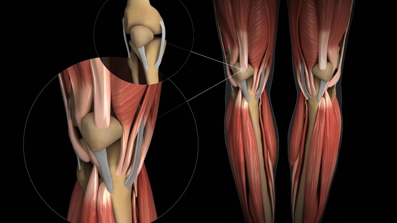
Parts and anatomy of the knee
The anatomical parts that belong to the knee are:
Bones and joints
The bones found in the knee are as follows:
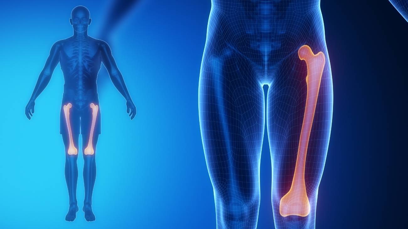
- Femur: It is the longest bone in the human body. It is joined (at its lower end) by means of the medial condyle and lateral condyle to the glenoid cavity of the tibia thanks to the cruciate and collateral ligaments. It lies posterior to the patella and attaches to the patella at the femoral trochlea.
- Patella: It is usually located in the centre of the knee and in the anterior area of the femoral trochlea. It is united with the quadriceps femoris tendon and, inferiorly, with the medial collateral ligament. It is a flattened and rounded bone, which contributes to joint movements.
- Tibia: The glenoid sockets on the upper surface of the tibia join with the femoral condyles to form the knee. In between are the menisci that help cushion shocks between the bones. It is attached to the femur and patella by tendons and ligaments.
The joints found in the knees are mentioned below:
- Lateral femorotibial: This joint is located at the lateral condyle of the femur and the glenoid socket of the tibia, it is responsible for extension movements of the knee.
- Medial femorotibial: Like the previous joint, this particular body gives movement to the femur, from its medial condyle to the flat surface of the tibia.
- Patellofemoral: The articular aspect of the patella is joined to the trochlea of the femur by this joint. It is in contact with the patellar ligaments and the tendons of the quadriceps femoris.

Muscles
The following muscles can be found in the knee:
- Rectus femoris: This tissue runs from the iliac spine on the inside and from the ilium to the tendon derived from the quadriceps femoris, forming part of this muscle group. Knee extension and hip flexion are actions that derive from this muscle.
- Vastus medialis: Located on the back of the leg, it is also part of the quadriceps. It can be called vastus internus because of the route it takes, from the trochanter of the femur to the patellar tendon. It allows extension of the knee.
- Vastus intermedius: This rectus muscle originates on the anterior surface of the femur, in the lateral area. It inserts into the patellar ligament, which allows it to work as an extensor of the leg.
- Vastus lateralis: The last muscle forming the quadriceps femoris group, it is also called vastus externus. Its action is knee extension and knee balance. It runs from the fibrous membrane to the trochanter of the femur.
- Popliteus: This tissue is located in the posterior area of the knee, allowing flexion and rotation of the knee. It arises from the external condyle of the femur and inserts into the glenoid sockets.
- Gastrocnemius: Also known as calf muscles. These muscles located at the back of the knee arise from the condyles of the femur to the calcaneal bone of the foot. Their mission is plantar flexion.
- Plantar: This is a thin muscle located at the back of the knee and runs above the gastrocnemius. It arises from the external condyle of the femur and inserts into the Achilles tendon, which allows flexion of the sole of the foot.
- The gracilis or internal rectus: It develops from the ischium and pubis to three different areas (symphysis pubis, medial condyle of the femur and medial condyle of the tibia). Its function is internal rotation, flexion and adduction of the hip.
- Sartorius: Its origin is in the anterior area of the thigh, in the iliac corner. It inserts on the upper surface of the tibia, which makes it the longest muscle in the body. In addition to being the hip flexor, it also allows flexion and extension of the knee.
- Biceps cruralis or femoris: It arises from the tuberosity of the ischium and the lateral lip of the femur to the upper area of the fibula. This allows it to be a leg flexor. It forms the ischiosural muscle group.
- Semitendinosus: This tissue generates the flexion of the leg and works as a hip extensor. It runs from the tuberosity of the ischium to the upper end of the tibia. It also belongs to the ischiosural muscles together with semimembranosus.
- Semimembranosus: The internal rotation of the knee is generated by the action of this muscle, which is also a hip extensor. It arises from the ischial tuberosity and inserts on the medial condyle of the tibia.
- Tensor of fascia lata: It arises from the spine of the ilium (anterosuperiorly) and develops to the iliotibial tendinous structure (in the lateral tubercle). Its action produces abduction, rotation and flexion of the hip. It is also a tensor of the fascia lata.

Ligaments and menisci
The connective tissue in the knee that binds this joint together are:
- Posterior cruciate: This connective tissue prevents slippage of the tibia with the femur. It runs from the lateral meniscus to the tibia in the intercondylar area. Surgery is one of the most effective treatments for repair.
- Anterior cruciate: Another of the main ligaments of the knee, its function is to prevent displacement of the femur and tibia. It arises from the lateral part of the femur to the medial area of the tibia.
- Tibial collateral: Also known as the lateral collateral ligament internally (LLI) because of its course. It arises from the internal condyle of the femur and inserts on the inner aspect of the tibia. Medial collateral is another name for it.
- Lateral external or lateral collateral (LLE): The femur is attached to the head of the fibula by means of this connective tissue. Its action is very important, as it prevents lateral displacement of the knee.
- Patellar: It is responsible for joining the lower part of the patella to the anterior tuberosity of the tibia. The extension of the leg is carried out in a balanced way thanks to this tissue.
- Transverse: Also known as the jugal ligament because it prevents the meniscus from moving internally.
- Posterior meniscofemoral: Its action is to allow the anterior meniscus to join the internal condyle of the femur.
- Anterior meniscofemoral: Some call it Humphrey's ligament after its discoverer and scholar. This connective tissue joins the internal condyle of the femur to the external meniscus, but passes in front of the posterior meniscofemoral meniscus.
- Popliteus obliquus: The external condyle of the femur is attached to the semimembranosus by this ligament.
- Popliteus arcuate: The external condyle of the femur also gives rise to this ligament up to the head of the fibula, creating balance in knee movements.
- Internal patellar meniscus: The action of this ligament is to allow the connection of the internal meniscus with the patella.
- External patellar meniscus: The patella and the external meniscus are joined by this ligament.
- Internal patellar alar: It is possible to keep the internal condyle of the femur together with the patella thanks to the function performed by this tape.
- External patellar alar: The external condyle of the femur and the patella, in its alar part, are connected through this tissue.
The menisci are cartilaginous tissues that lie between the femur and the tibia. We will show you each of them below:
- Internal: Its shape is C-shaped, its function is to cushion the blows between the bones of the knee and to cause a perfect union between both bony tissues. This is due to the low concavity of the glenoid cavities and the greater convexity of the femoral condyles.
- External: In this case it is O-shaped and is also free of nerve and blood vessel endings. The internal part remains loose in order to reduce the impact of movements between the bones.

Best products for knee and leg pain relief
Bestseller
-

2 Knee Compression Sleeve (Black/Gray)
£17,50 -
2 Knee Compression Sleeve (Green/Navy)
£17,50 -
2 Knee Compression Sleeve (Pink/Bordeaux)
£17,50 -
2 Patella Knee Strap (Black/Gray)
£12,95 -
2 Patella Knee Strap (Green/Navy)
£12,95 -
2 Patella Knee Strap (Pink/Bordeaux)
£12,95 -

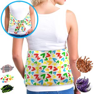
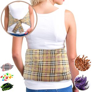
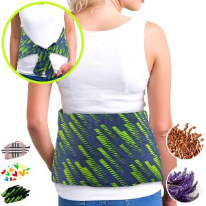
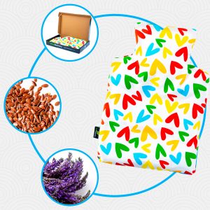
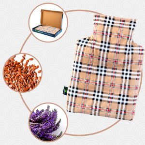
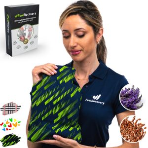
Microwave Wheat Bag for Back Pain Relief (Extra Large) (Hearts)
£25,50 -
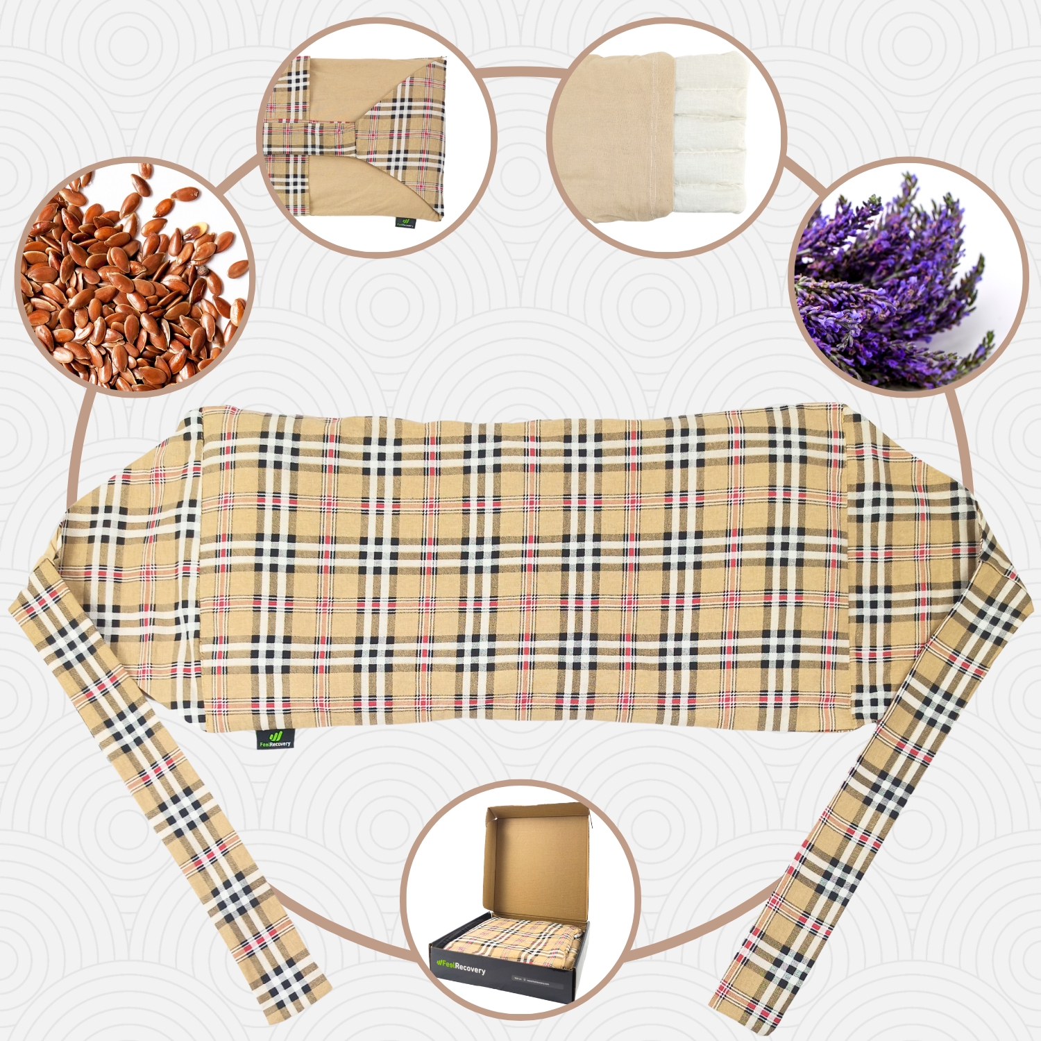
Microwave Wheat Bag for Back Pain Relief (Extra Large) (Oxford)
£25,50 -
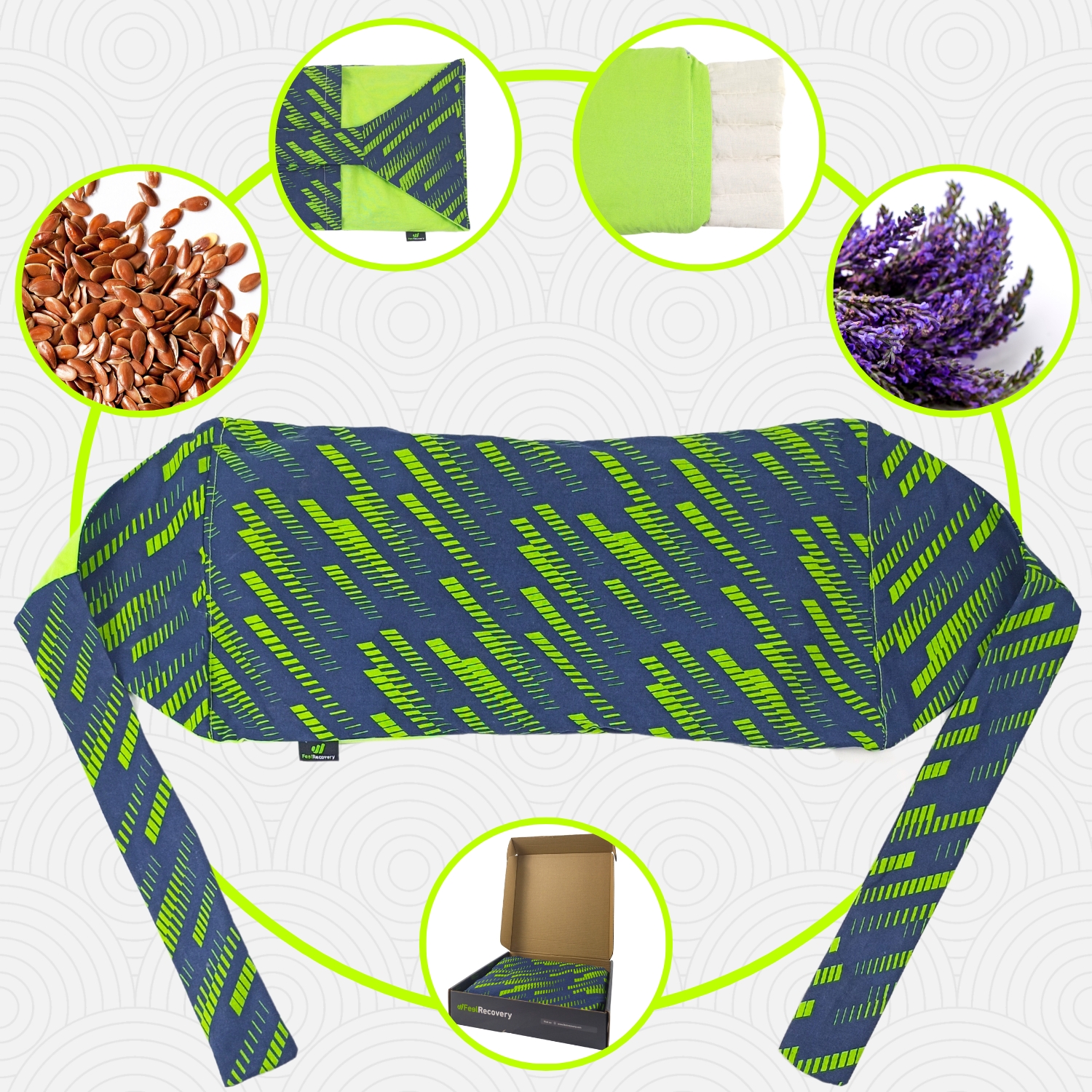
Microwave Wheat Bag for Back Pain Relief (Extra Large) (Sport)
£25,50 -
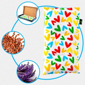
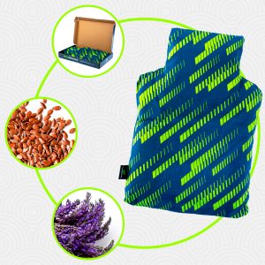
Microwaveable Wheat Bag for Pain Relief (Hearts)
£17,50 -
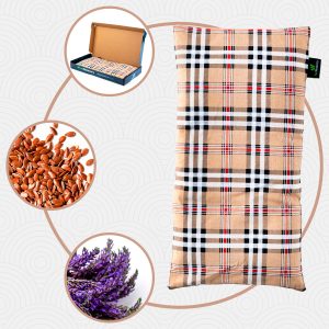
Microwaveable Wheat Bag for Pain Relief (Oxford)
£17,50 -
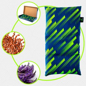
Microwaveable Wheat Bag for Pain Relief (Sport)
£17,50 -
Reusable Gel Ice Packs for Knees with Compression Band
£17,50 -
Reusable Gel Ice Packs with Compression Band for Sports Injury
£17,50 -
Wheat Bag for Microwave Classic Bottle Shaped (Hearts)
£17,50 -
Wheat Bag for Microwave Classic Bottle Shaped (Oxford)
£17,50 -
Wheat Bag for Microwave Classic Bottle Shaped (Sport)
£17,50


Biomechanics of the knee
The knee can perform the following biomechanical movements:
- Flexion: This action involves placing the heel as close as possible to the gluteal area and can have an opening angle of 130° to 170° thanks to the work of the hamstrings, the sartorius and the popliteus.
- Extension: This movement is the opposite of flexion and is performed when the tibia and fibula are in a straight line with the femur. It is a resting movement in which the opening is 0°.
- Internal rotation: This consists of turning the foot towards the inside of the body, with the tibia as the axis. The opening can be up to 40° or 50°, the cruciate ligaments work to prevent tibial displacement.
- External rotation: This is a biomechanical action performed by the knee when the tibia is taken as the axis and the foot is rotated to the collateral side. The patellar ligament, meniscus and cruciate ligaments are important for this task.
Most common knee injuries
There are different diseases that occur in the knee, so we will explain below which are the most common ones.
Types of knee injuries
The most common types of knee injuries are:
- Knee osteoarthritis: This is a degenerative disease that causes wear and tear on the cartilage of the patellofemoral and patellofemoral joints. This causes the bones to contact each other during movement, resulting in severe pain, swelling and joint stiffness. Its causes are varied, but age and demanding actions are the most common. It is also called gonarthrosis.
- Bursitis of the knee: There are several serous bursae in the knee that contain synovial fluid to improve cushioning during joint movements. When these bursae become inflamed due to a blow or excessive strain, among other reasons, this is called "knee bursitis". The most common is patellar bursitis.
- Knee sprain: It is possible to find different degrees of these injuries that occur when, due to a bad movement or trauma, the ligaments (especially the four most important ones) are partially or completely torn. This causes a decrease in movement and instability when walking.
- Chondromalacia patellae: The articular surface of the patella is affected by this syndrome which causes pain when the knee is used. It is one of the main injuries of this joint caused by the wear and tear of the cartilage when performing demanding activities or bearing too much weight.
- Patellar tendonitis of the knee: Inflammation in the patellar tendon can occur for a variety of reasons, the most common being sports activities and advanced age. Common symptoms include pain behind the kneecap and redness.
- Knee joint dislocation: Is the separation of the lower part of the femur, the condyles, from the upper part of the tibia, the glenoid sockets. This separation of the bones can be complete or partial, the latter being called knee subluxation.
- Kneecap dislocation: The patella is usually located almost in the centre of the knee, supported at the anterior superior part by the quadriceps femoris tendon and by the patellar ligament at the other end. In turn, the lateral and medial ligaments provide the force necessary to hold this flat bone in its natural position.
Sports injuries to the knees
The most common ailments that occur in the knee from playing different sports are
- Knee injuries in badminton: Knee injuries are the most common injuries suffered by badminton athletes. Repetitive and high-impact movements cause injuries to the cruciate and lateral ligaments, as well as patellar tendonitis and chondromalacia.
- Knee injuries in basketball: Patellar dislocation, meniscus wear, lateral and cruciate ligament sprains, patellar tendonitis and inflammation in the quadriceps tendon are common ailments that these athletes have. In some cases osteoarthritis of the knee may occur.
- Knee and leg injuries in cycling: Knee contusions in cyclists are frequent due to the high demand on the patellar tendon, the biceps cruris, the tensor fascia lata and popliteus. Therefore, patellar bursitis, tendinopathies and muscle contracture is common in these parts.
- Knee injuries in Crossfit: Wear and tear of the meniscus, erosion of the articular cartilage, inflammation of the patellar synovial bursa and the quadriceps tendon are the most common injuries that occur in this activity. Ligament injuries can also be encountered.
- Knee injuries in climbing: In climbing, the connective tissues of the knee are the most strained, so sprains are common. Knee flexion can also lead to inflammation of the patellar bursa and gastrocnemius contractures.
- Knee injuries in football: The cruciate and collateral ligaments are the ligamentous tissues that suffer most. It is also common to find meniscus tears, hamstring and calf contractures, patellar bursitis and wear and tear on the cartilage of the femoral condyle.
- Knee injuries in golf: The lateral collateral, medial collateral and cruciate are the connective tissues that suffer the most injuries. On the other hand, the articular cartilage suffers significant wear and tear, which can lead to osteoarthritis. The quadriceps tendon is also prone to injury.
- Knee injuries in weightlifting: The practice of this activity makes the knee support an excessive amount of weight, so patellar bursitis, inflammation of the tendons and rupture of cruciate ligaments are frequent. Dislocations are also common in the sport.
- Knee injuries in rugby: Cruciate ligament tears, inflammation of the hamstring and patellar bursitis are the three most common types of injuries in rugby. Patella fractures, tendonitis and muscle contractures are also possible.
- Knee injuries in running: The lateral and collateral ligaments are the tissues that suffer most in this activity to prevent the tibia from sliding in relation to the femur. It is possible to find athletes with chondromalacia patellae, calf, hamstring and quadriceps femoris contractures.
- Knee injuries in tennis: The movements practised in this sport cause injuries to both menisci, inflammation in the patellar tendon, chondropathy patellofemoral, contractions in the quadriceps and hamstring dislocations.
Diseases and ailments in the knees

There are other types of ailments that can appear in the knees, which are:
Osteochondrosis
This is also known as Osgood-Schlatter disease. This is the appearance of a lump in the front part of the knee, causing inflammation and pain. It occurs particularly at the time of bone growth, making children and young people the most vulnerable age group.
Lupus erythematosus
Lupus erythematosus is a disease caused by the body's own immune system that causes wear and tear on the articular cartilage of the knee. This leads to inflammation and stiffness of movement in this part of the body.
Hoffa's syndrome
The pain and swelling in the knee is caused by swelling that affects the deep area at the junction of the patella and patellar tendon. This fatty area is frequently injured in young people and athletes due to the demanding activities the joint endures.
Baker's cyst
This knee condition is caused by excessive accumulation of synovial fluid, forming a cyst that presses on the back of the joint. This causes severe pain and joint immobility in the patient.
Knee hyperextension
This ailment is known as Genu Recurvatum, which consists of keeping the knee extended more than 10° due to a malfunction of the cruciate ligaments. It can also be caused by rickets or poliomyelitis.
How can we relieve knee pain through complementary and non-invasive therapies?
Take a look at the following complementary and non-invasive therapies to help improve knee pain symptoms:
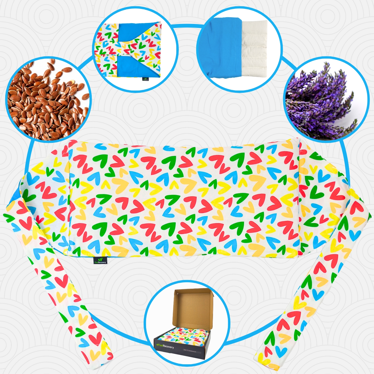
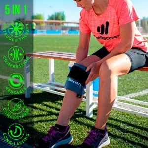
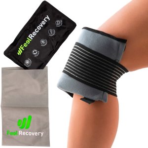
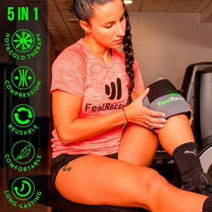
Heat and cold therapy
This non-invasive therapy involves the application of gels, heat packs, warm water or ice packs that produce heat and cold to the affected area. In this way, pain and inflammation are reduced much more quickly. To do this, it will be necessary to apply both temperatures for 5 minutes each, but bearing in mind that the session should not exceed 15 to 20 minutes. You should start and end with heat for best results.
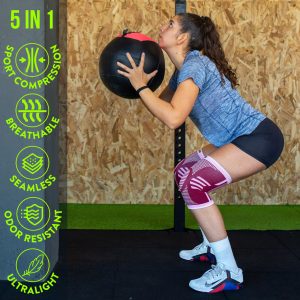
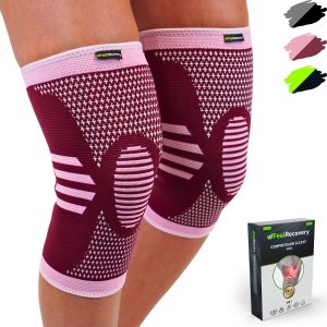
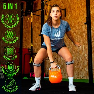
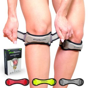
Compression therapy
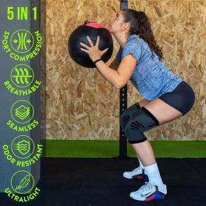
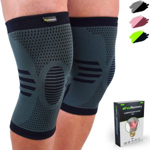
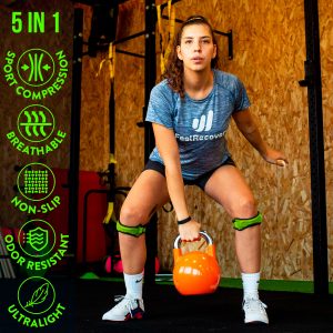
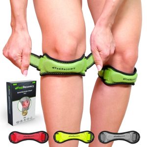
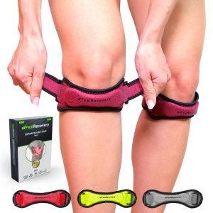
It is possible to apply this therapy to the knee to prevent further injury and to produce gas exchange between the blood and the tissues. To get the disease into remission as quickly as possible, the application of compression sportswear, patellar tendonitis tape and compression sports knee braces is necessary. These items will improve blood circulation and decrease pain more effectively.
Massage therapy
Massage rollers and massage guns are useful items that can be used in this non-invasive complementary therapy to reduce the symptoms of knee pain and swelling. It is necessary to consult your doctor beforehand so that you can choose the most suitable item for you, as blood stimulation is different for each case.
Acupressure therapy
Acupressure is an ancient technique that is very effective in eliminating the symptoms of patellar pain by massaging strategic areas. This enhances the dilation of the capillary walls to allow a reduction in the levels of toxic molecules in the fibres. This non-invasive therapy can be done through the use of different products; for example, hard or soft massage balls, acupuncture hooks and foot massagers.
Thermotherapy
There are different techniques that help to reduce pain in the knees, one of the most efficient being thermotherapy. This is due to the benefits that heat produces when applied directly to the affected area. For this reason, it is advisable to consult a doctor before choosing this non-invasive treatment and to be able to use thermal heat pillows that improve the dilation of the capillary walls.
Cryotherapy
Cryotherapy can be used for pain and inflammation caused by patellar tendonitis and other injuries. For this purpose, cold should be used directly on the affected area two to three times per day, depending on the discretion of the treating physician. It is possible to use ice gel packs to obtain a pleasant and efficient temperature.
Electrical muscle stimulation (EMS)
Muscle electrostimulation, or EMS, is a therapy that consists of stimulating muscle contractions through the use of electricity, so as to achieve an effect of activity and hypertrophy as in the gym, but without the need to go to any sports center. This means that you can put your muscles to work without leaving home.
Electrotherapy
This is a technique that seeks relief from pain and some physical ailments through the application of electrical and electromagnetic energy, among other variants, through the skin with the use of conductive pads called electrodes. It is a very safe type of therapy and must be applied by a physical therapist specialized in the manipulation of electricity to treat some kinds of ailments.
Myofascial release therapy
Also known as myofascial induction, this therapy consists of the application of manual massage to treat the shortening and tension generated in the myofascial tissue that connects the muscles to the bones and nerves. For this purpose, various massage techniques are used that focus on the so-called trigger points.
Percussion Massage Therapy
Vibration or percussion massages are precise, rhythmic and energetic strokes on the body to achieve relief from some annoying symptoms when muscle fibers are tightened, often by a high workload on them and that has left trigger points in the muscle fibers.
R.I.C.E Therapy
The R.I.C.E. therapy is the first and simplest of the treatment protocols for minor injuries. It appears in the sports field to deal with accidents involving acute injuries. For many years, it has been considered the most suitable for its speed and results.
Trigger points therapy
Myofascial pain points or trigger points are knots that are created in the deeper muscle tissues, causing intense pain. The pain does not always manifest itself right in the area where the point develops, but rather this pain is referred to nearby areas that seemingly do not appear to be related. In fact, it is estimated that more than 80% of the pain they cause manifests in other parts of the body.
Other effective alternative therapies
It is also possible to treat knee pain with other non-invasive therapies, which are mentioned below:
- Natural remedies using plants: Plants create soothing effects and stimulate the blood supply to reduce pain. This is why phytotherapy is used for knee injuries. It can be delivered by infusions or baths of lemon balm, boldo or lime blossom.
- Acupuncture: Knee pain can be reduced by means of this oriental medicine, which consists of stimulating the nervous system so that the patient feels less pain.
- Kinesiotherapy: This is a complementary therapy that seeks to improve knee movements by means of specific exercises that are short and repetitive. It is advisable to consult a doctor beforehand to avoid future injuries.
- Aromatherapy: The use of concentrated liquids with aromas of lemon, boldo, linden and ginger helps the patient to better manage their illness or knee ailment. This is one of the least invasive techniques available to combat patellar pain.
References
- Blackburn, T. A., & Craig, E. (1980). Knee anatomy: a brief review. Physical therapy, 60(12), 1556-1560. https://academic.oup.com/ptj/article-abstract/60/12/1556/2727156?login=false
- Abulhasan, J. F., & Grey, M. J. (2017). Anatomy and physiology of knee stability. Journal of Functional Morphology and kinesiology, 2(4), 34. https://www.mdpi.com/2411-5142/2/4/34
- Flandry, F., & Hommel, G. (2011). Normal anatomy and biomechanics of the knee. Sports medicine and arthroscopy review, 19(2), 82-92. https://journals.lww.com/sportsmedarthro/Abstract/2011/06000/Normal_Anatomy_and_Biomechanics_of_the_Knee.2.aspx
- Fulkerson, J. P., & Gossling, H. R. (1980). Anatomy of the knee joint lateral retinaculum. Clin Orthop Relat Res, 153, 183-188. https://www.researchgate.net/profile/John-Fulkerson/publication/246367558_Insertion_orientation_of_terminal_vastus_lateralis_obliquus_and_vastus_medialis_obliquus_muscle_fibers_in_human_knees/links/5ce13498a6fdccc9ddbc7cc9/Insertion-orientation-of-terminal-vastus-lateralis-obliquus-and-vastus-medialis-obliquus-muscle-fibers-in-human-knees.pdf
- Roos, E. M. (2005). Joint injury causes knee osteoarthritis in young adults. Current opinion in rheumatology, 17(2), 195-200. https://journals.lww.com/co-rheumatology/Abstract/2005/03000/Joint_injury_causes_knee_osteoarthritis_in_young.16.aspx
- Smillie, I. S. (1946). Injuries of the knee joint. Annexe Thesis Digitisation Project 2019 Block 22. https://era.ed.ac.uk/handle/1842/33992
- Abbott, L. C., John, B., Saunders, M., Bost, F. C., & Anderson, C. E. (1944). Injuries to the ligaments of the knee joint. JBJS, 26(3), 503-521. https://journals.lww.com/jbjsjournal/Abstract/1944/26030/INJURIES_TO_THE_LIGAMENTS_OF_THE_KNEE_JOINT.11.aspx
- Gelber, A. C., Hochberg, M. C., Mead, L. A., Wang, N. Y., Wigley, F. M., & Klag, M. J. (2000). Joint injury in young adults and risk for subsequent knee and hip osteoarthritis. Annals of internal medicine, 133(5), 321-328. https://www.acpjournals.org/doi/abs/10.7326/0003-4819-133-5-200009050-00007
- Arden, N. K., Crozier, S., Smith, H., Anderson, F., Edwards, C., Raphael, H., & Cooper, C. (2006). Knee pain, knee osteoarthritis, and the risk of fracture. Arthritis Care & Research: Official Journal of the American College of Rheumatology, 55(4), 610-615. https://onlinelibrary.wiley.com/doi/abs/10.1002/art.22088
- Barrack, R. L., Schrader, T., Bertot, A. J., Wolfe, M. W., & Myers, L. (2001). Component rotation and anterior knee pain after total knee arthroplasty. Clinical Orthopaedics and Related Research (1976-2007), 392, 46-55. https://journals.lww.com/corr/Abstract/2001/11000/Component_Rotation_and_Anterior_Knee_Pain_After.6.aspx