- Definition: What is a bone fracture or broken bone?
- What are the most common types of fractures?
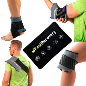
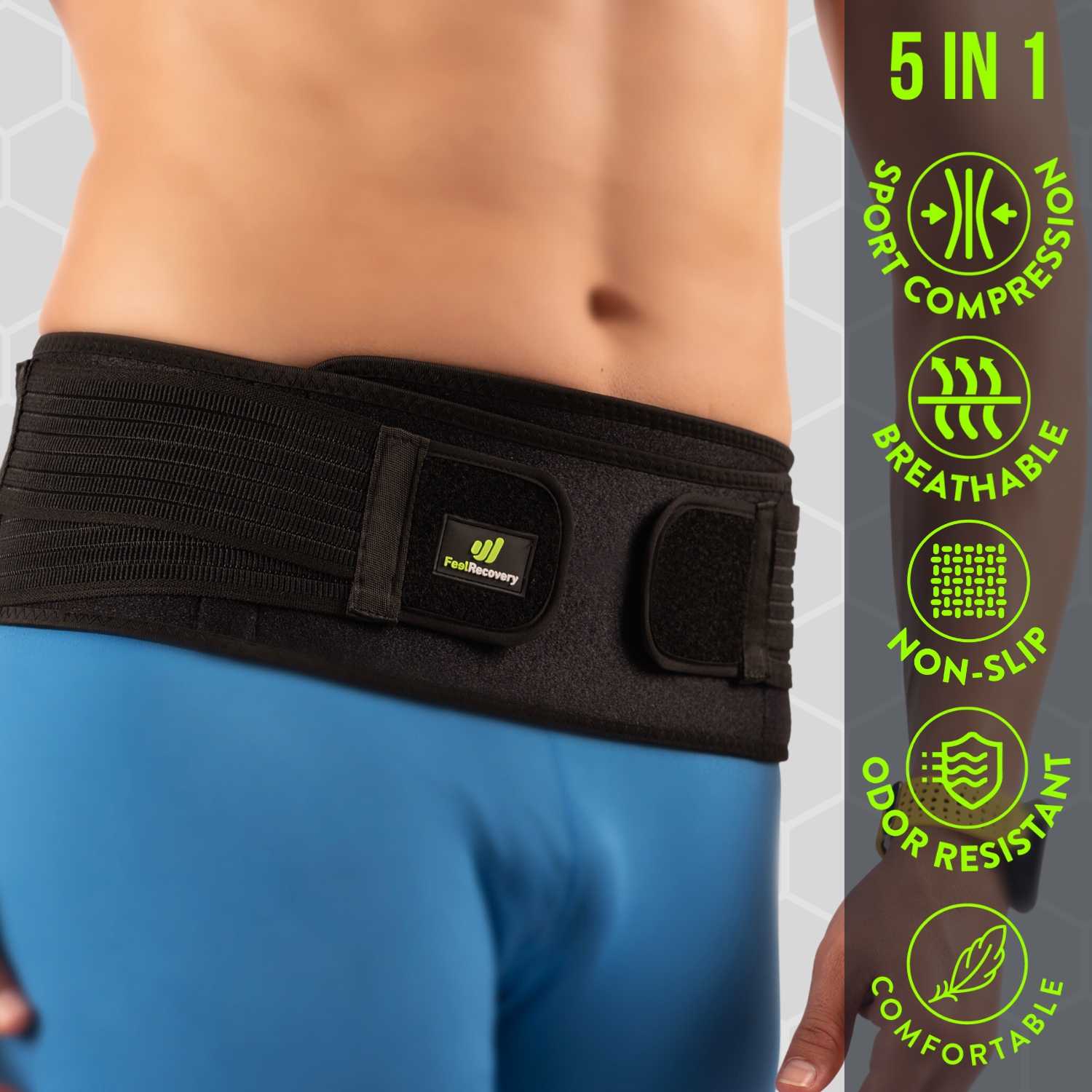
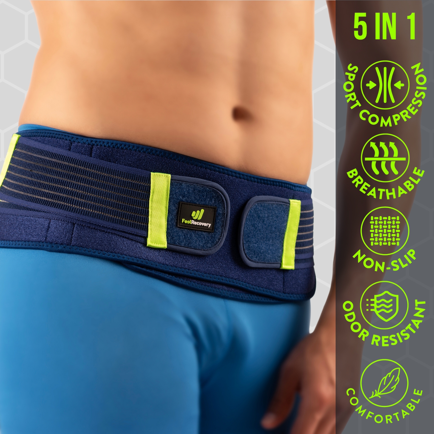
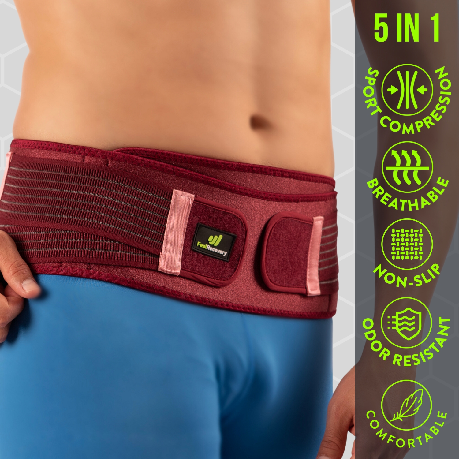
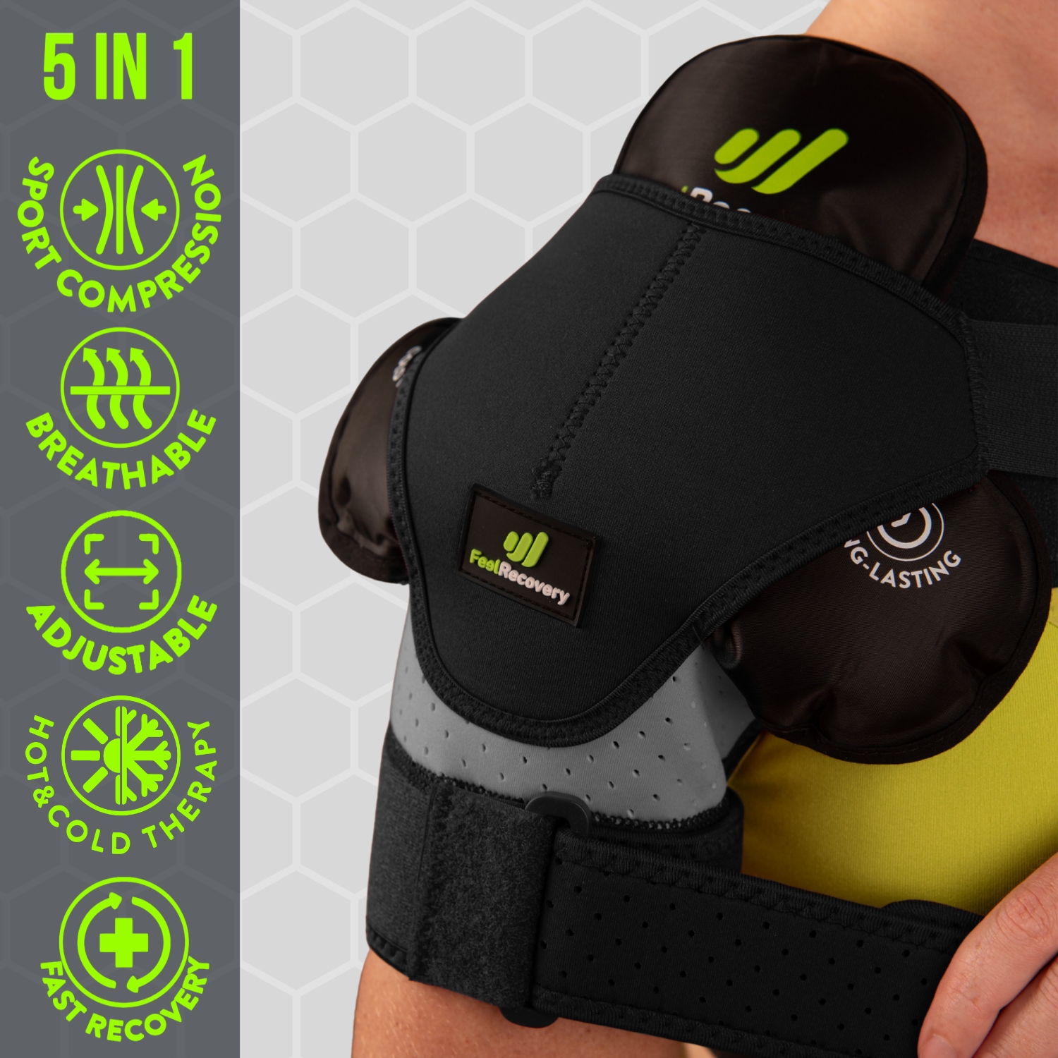
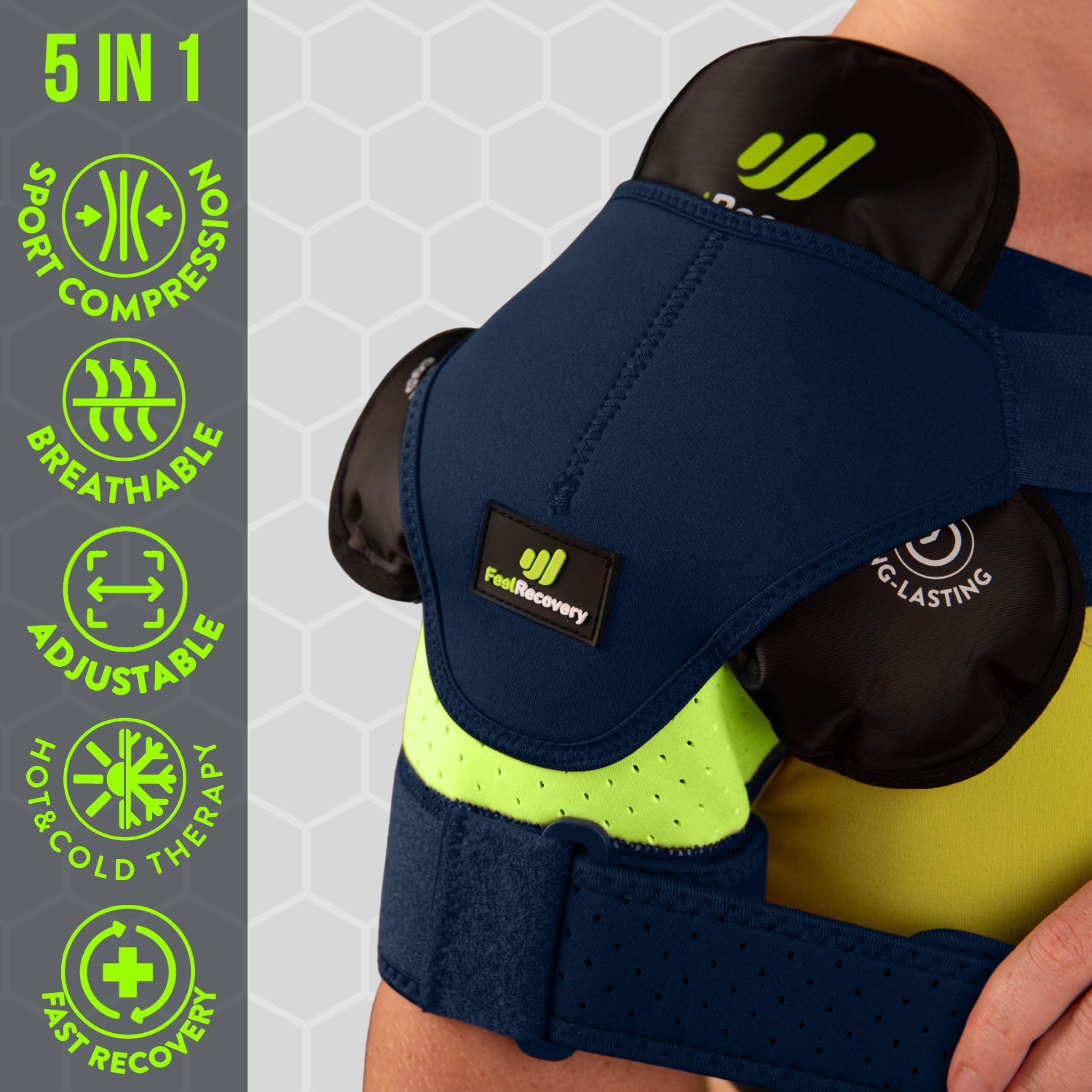
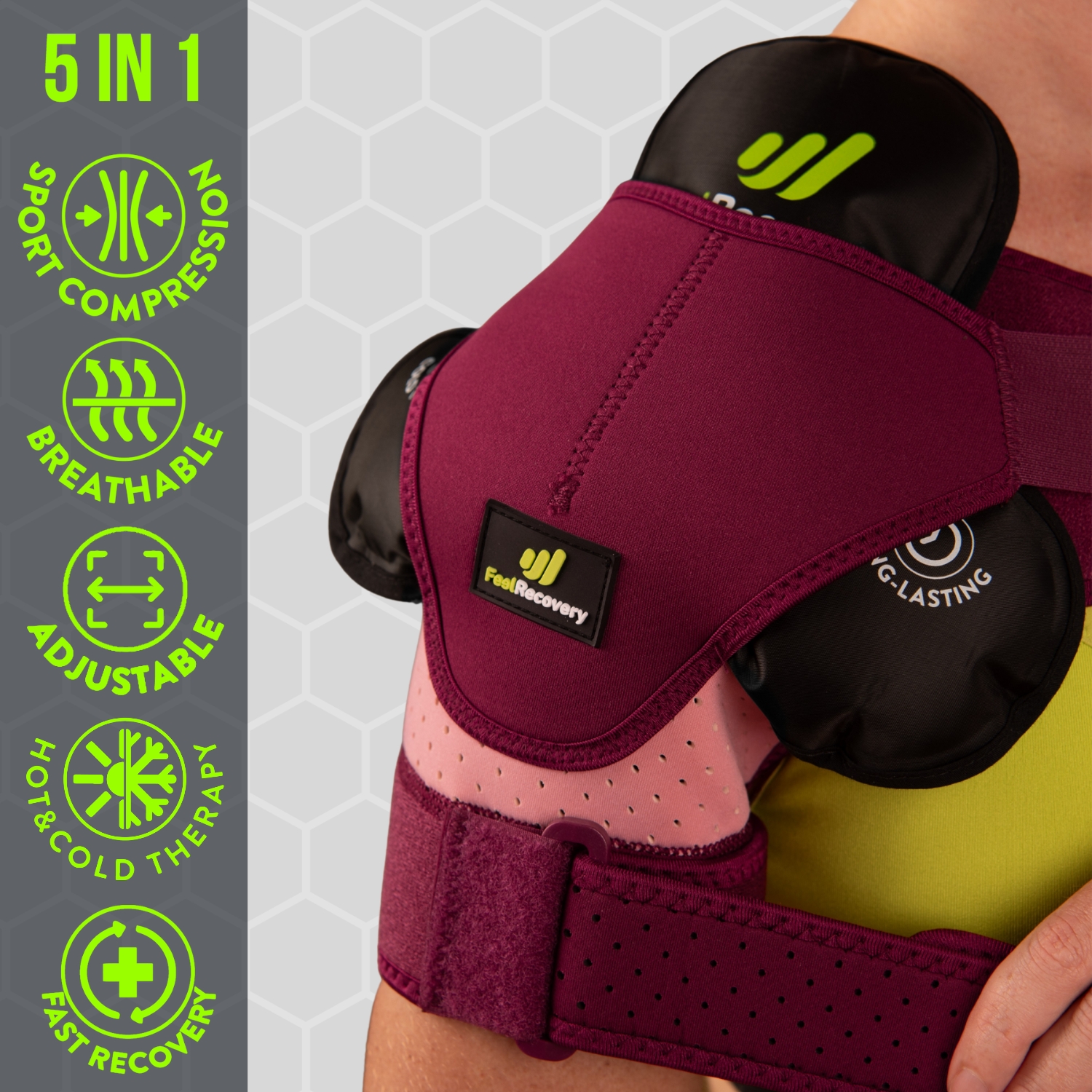
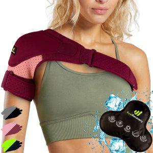
- Best bone fractures products
- What are the causes and risk factors for bone fractures?
- What is the first aid care for treating a bone fracture?
- What methods of prevention to avoid broken bones are the most effective?
- F.A.Q: Frequently asked questions
All tissues in the human body have the ability to regenerate after injury. By nature, the body has the ability to produce the cells and substances necessary to rebuild the tissues immediately after the contusion. As a result, it provides the same characteristics to the injured tissue before its laceration.
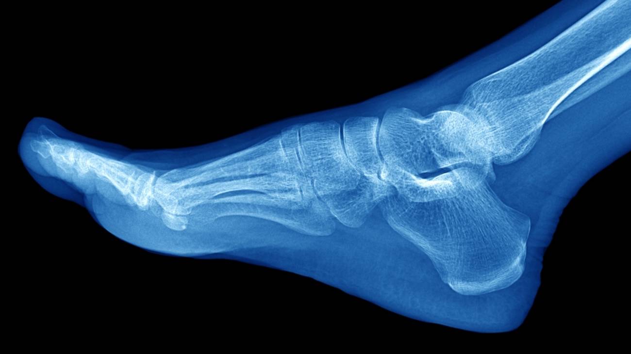
One of the strongest tissues of all is bone. But although these layers are hard and tough, they have the potential to break down. If they do, a bone fracture occurs, triggering various symptoms. So, here you will find out what they are and what the main types are.
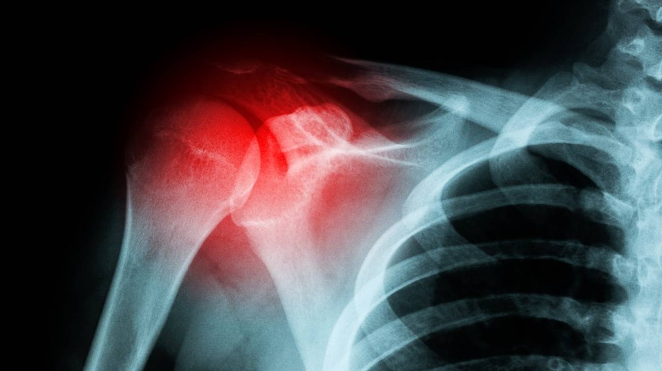
Definition: What is a bone fracture or broken bone?
A bone fracture is defined as the cracking, partial or total breakage of a bone throughout the human body. This is the result of sports injuries, sudden falls, sudden accidents or overuse of a bony area belonging to a person's skeletal system.
Since the body is made up of 206 bones and these are responsible for providing support, protecting internal organs and allowing movement, a fracture tends to trigger considerable signs and symptoms in patients. They are mainly characterised by intense pain, swelling or bruising, deformity of the limb, loss or decrease of sensation and complications in terms of mobility.
However, in order to diagnose a fracture on a medical level and define its state of seriousness, apart from assessing the symptoms of the affected person, the health professional must also study the reasons for the injury and carry out a clinical examination to obtain more precise results. Under this, it will be possible to establish a personalised treatment that will speed up the rehabilitation and recovery process, according to all the references of the breakage of a given bone.
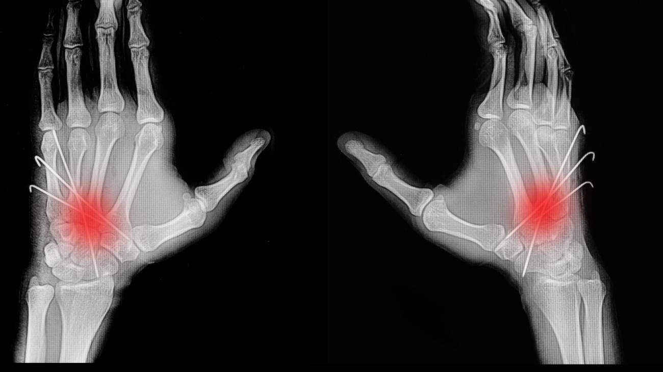
What are the most common types of fractures?

You probably didn't know this yet, but the truth is that there is no single type of fracture. Depending on the type of damage that manifests the injury.
These bone breaks are classified as follows in order to provide a more specific diagnosis:
- Complete fracture: This is the most common type of fracture or break. Basically, it is distinguished by breaking the bone into two parts and, therefore, the fracture line affects the bone itself around its entire circumference and divides it into two fragments. Sometimes, it can be assessed as a single fracture.
- Simple fracture: This is usually the result of a severe injury. The fracture involves a single fracture line through the affected bone and may also be identified as a transverse, oblique and spiral fracture.
- Open or exposed fracture: This is an injury in which there is communication between the impacted bone and the exterior, due to a concurrent fracture of the skin and soft tissues that protect the focus of the fracture (a wound or haematoma). Consequently, the broken bone protrudes through the patient's skin and therefore, in this type of fracture, there is a potential for infections to develop from bacterial flora on the skin and/or in the environment.
- Closed fracture: Unlike the previous type of break, this is defined as an injury in which the bone does not protrude through the skin, even though there is a break in the bone. Since the skin and tissues covering the fractured bone are not injured by the fracture, but the pain it generates makes it possible to identify this type of fracture. It usually occurs in the femur, hip, ankle, wrist, tibia and fibula.
- Transverse fracture: This refers to those fractures that present a perpendicular trajectory to the major axis of the bone, so that the bone fragments are not deviated. Apart from this, they differ in that they are easy to reduce (i.e. they allow the bone fragments to adjust quickly) and are stable (i.e. the bone portions do not tend to move). Consequently, their prognosis is favourable and their treatment is rapid.
- Oblique fracture without displacement: As its name suggests, it causes the bone to break at an angle. Moreover, as there is no displacement, it means that the impacted ends are separated from each other. In most cases, this specific pathology is triggered by direct and indirect trauma (heavy weight on the bone or due to some bending mechanism, respectively).
- Oblique fracture with displacement: It also develops an oblique tear, but in this case, separation of the ends of the lesion is observed. For this reason, the complexity of the fracture is greater, in view of the fact that it has a negative influence on the tissues around the fracture. For this reason, its treatment is more complicated than that required for an oblique fracture without displacement, due to the instability it exhibits.
- Butterfly wing fracture: Specifically, it is defined as a fracture that presents an intermediate wedge-shaped fragment, once the injury is manifested. Thus, it consists of a typical trace when bending forces occur on the bone being breached.
- Spiral fracture: Also known as "spiroid", these are defined as conditions in which the angle of the break crosses the bone diagonally and are therefore similar to oblique fractures. However, to differentiate them, they develop a rotational element that slides into the bone longitudinally. They are usually caused by torsional forces and the trace presents a spiral around the bony portion. It should be noted that they are considered to be rare.
- Segmental fracture: In this case, the afflicted bone segment remains isolated from the ends, in its entirety. Therefore, the greatest risk associated with this type of injury is the loss or excessive reduction of blood supply to the affected area.
- Comminuted fracture: This is based on a pathology that involves the fracture of the bone into multiple fragments (three or more parts) and because of this, it is also called a "multifragmentary fracture". For this reason, it is similar to a broken glass that spreads many pieces of itself. Consequently, it is one of the most serious of all fractures and is usually caused by trauma.
- Parcel fracture: These are fractures that result in the breakage of non-essential or structural parts of the impacted bone. Such as, for example, avulsions that are generated once a muscular force tears off the part of the bone in which the affiliated muscle is anchored.
- Supracondylar fractures: In most cases, these are injuries that occur in patients under 8 years of age. They are located above the elbow and tend to break the arm bone or humerus at a short distance from the upper elbow. Among the most frequent causes are traffic accidents, sports contusions and direct blows. In case it reaches its highest level of severity, it is possible to trigger circulation disorder and nerve damage.
- Stress fracture: Stress fractures are fractures that develop when repetitive pressure is placed on bones. Since, this type of movement usually weakens the bone structure over time, until it causes an injury leading to the fracture. In general, the main patients of this disease are running and jumping athletes, women with the female athlete triad and military personnel.
- Impacted fracture: This is a type of injury that is identified as one bone fragment impinging on another. This means that the bone portions are compressed into each other and is almost always caused by compressive force (especially in regions of cancellous bone).
- Green stem fracture: If a green stem fracture is diagnosed, it means that the bone has broken without separating into two parts. Thus, since children have a high elasticity in their bones that tends to splinter the bony part easily, these fractures are typical for children. However, they are distinguished by their simple reduction, as there is no displacement.
- Pathological fractures: As the name suggests, these are broken bones that develop for pathological reasons in a particular patient. Either because they suffer from osteoporosis, tumours or bone cancer, and naturally, these conditions favour the weakness of the bones so that they break easily (even when faced with light forces).
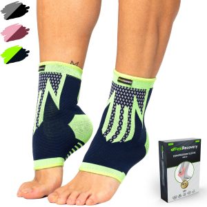
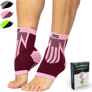
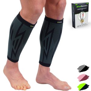
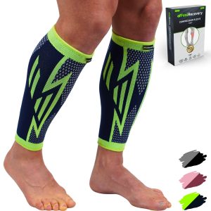
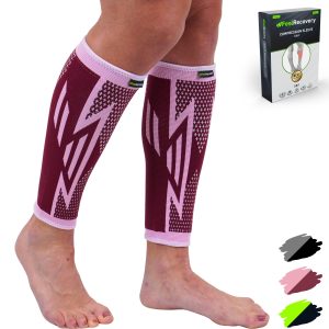
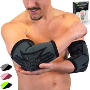
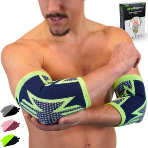
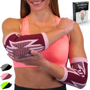
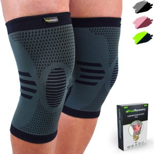
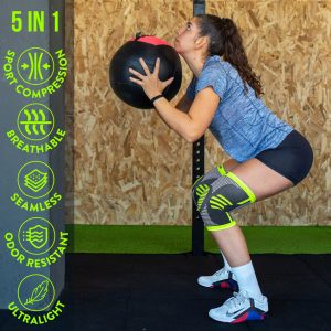
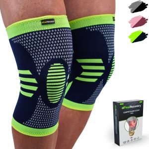
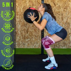
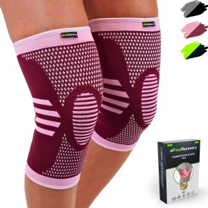
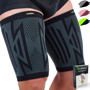
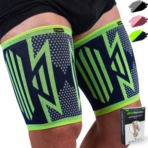
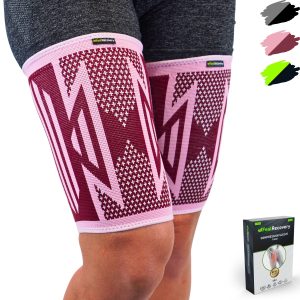
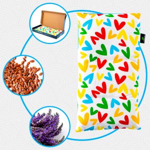
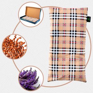
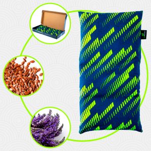
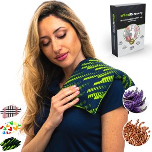
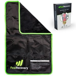
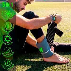
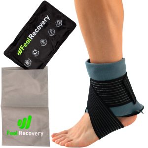
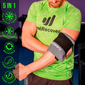
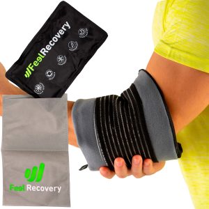
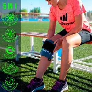
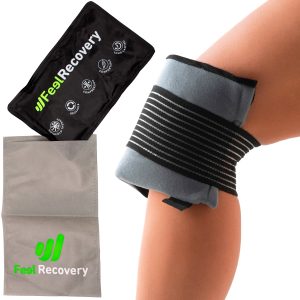
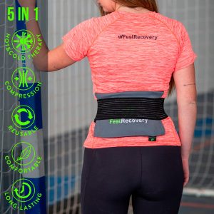
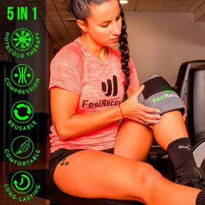
Best bone fractures products
Bestseller
-

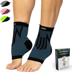
2 Ankle Compression Sleeve (Black/Gray)
$19.95 -
2 Ankle Compression Sleeve (Green/Navy)
$19.95 -
2 Ankle Compression Sleeve (Pink/Bordeaux)
$19.95 -
2 Calf Compression Sleeve (Black/Gray)
$19.95 -
2 Calf Compression Sleeve (Green/Navy)
$19.95 -
2 Calf Compression Sleeve (Pink/Bordeaux)
$19.95 -
2 Elbow Compression Sleeve (Black/Gray)
$19.95 -
2 Elbow Compression Sleeve (Green/Navy)
$19.95 -
2 Elbow Compression Sleeve (Pink/Bordeaux)
$19.95 -
2 Knee Compression Sleeve (Black/Gray)
$19.95 -
2 Knee Compression Sleeve (Green/Navy)
$19.95 -
2 Knee Compression Sleeve (Pink/Bordeaux)
$19.95 -
2 Thigh Compression Sleeve (Black/Gray)
$19.95 -
2 Thigh Compression Sleeve (Green/Navy)
$19.95 -
2 Thigh Compression Sleeve (Pink/Bordeaux)
$19.95 -
Microwaveable Heating Pad for Pain Relief (Hearts)
$19.95 -
Microwaveable Heating Pad for Pain Relief (Oxford)
$19.95 -
Microwaveable Heating Pad for Pain Relief (Sport)
$19.95 -
Reusable Extra Large Ice Packs for Back & Legs Pain
$29.95 -
Reusable Gel Ice Packs for Ankle & Foot with Compression Band
$19.95 -
Reusable Gel Ice Packs for Elbow & Arms with Compression Band
$19.95 -
Reusable Gel Ice Packs for Knees with Compression Band
$19.95 -
Reusable Gel Ice Packs for Shoulder & Back with Compression Band
$19.95 -
Reusable Gel Ice Packs with Compression Band for Sports Injury
$19.95 -

Sacroiliac Support Belt (Black)
$24.95 -
Sacroiliac Support Belt (Green)
$24.95 -
Sacroiliac Support Belt (Pink)
$24.95 -
Shoulder Support Brace (Black)
$24.95 -
Shoulder Support Brace (Green)
$24.95 -
Shoulder Support Brace (Pink)
$24.95 -
Wrist Brace (Black/Gray)
$19.95 -
Wrist Brace (Green/Navy)
$19.95 -
Wrist Brace (Pink/Bordeaux)
$19.95


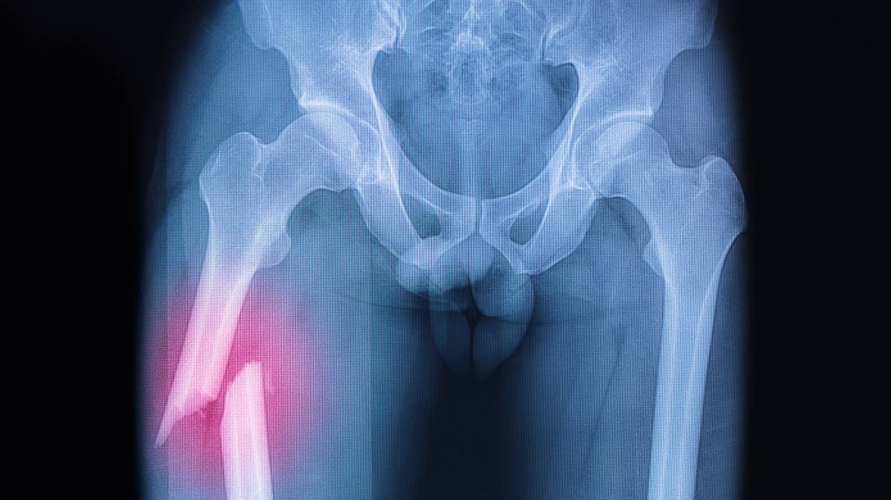
What are the causes and risk factors for bone fractures?
In most cases, bone fractures are caused when high pressure is applied to a bone and as a result, the bone breaks.
Below, we highlight the most common reasons that promote the occurrence of a bone fracture:
- Direct trauma in which the energy is transmitted between the skin and the soft parts or on the fractured point. For example: direct blows or sudden falls.
- Indirect trauma also causes a fracture. In this case, the injury is the result of a contusion formed at a certain distance from the force that generates it. For example, when a person suffers a sudden eventuality and falls to the ground on his hand, but breaks the bones in his shoulder. Or when an athlete makes a certain rotation of the leg that produces a fracture at the medial level of the fibula and tibia.
- The excessive or frequent use of a bone, as well as the intensity applied to it when carrying out certain repetitive actions, is another of the most common causes of a fracture. In this case, they are caused by fatigue or stress.
- Some diseases that support bone weakening, such as osteoporosis, bone tumours, bone cancer, etc. are considered to be strong risk factors for experiencing fractures of this type.
- If an individual is involved in a car accident, it is possible that he or she may suffer from one or more fractures in his or her body. The sudden forces of the collision tend to break bone structures very easily.
- Another reason why a person may experience a fracture is physical abuse. Either by another individual or by performing various high-risk sporting activities.
- When bones are subjected to forces to which they are not accustomed, without sufficient time for recovery and resorption of cells, it is very likely that such an injury will be triggered. In this case, it is identified as a stress fracture.
What is the first aid care for treating a bone fracture?

In order to apply appropriate first aid care after a bone fracture, it is first of all important to determine that the patient has suffered an injury.
By unqualified personnel
Whereas, some signs to deduce this are as follows:
- The injured person is unable to move the fractured region.
- The pain is intense and increases with touch.
- The discomfort is persistent for a prolonged period of time.
- The injured area is swollen.
- Ecchymosis or deformity and/or there is a skin wound (through which the bone protrudes).
Now, when detecting or assuming that it is a bone fracture, it is paramount that the patient and the people trying to help him/her help to implement the following, in order to prevent the injury from worsening:
- Holding the broken bone in a fixed and firm position. In other words, it should innately immobilise the injured part to prevent it from shifting and hurting further.
- In case the bony portion is protruding, just cover the wound with a clean towel or cloth. It is not advisable to try to fit or insert it.
- If there is bleeding from the fractured part, it is best to control it by applying pressure with a clean cloth to prevent the injured person from bleeding.
- If the visualised bleeding is coming from the nose, mouth or ears, it is not appropriate to stop the bleeding.
- If the fracture has impacted the head area, the patient should try to keep the head slightly higher than the rest of the body and avoid moving it. In addition, every effort should be made to keep the person from falling asleep.
- Seek medical help and call for help as quickly as possible, in order to treat the fracture quickly.
Once the patient is under medical supervision, the intervening orthopaedic surgeon or specialist in the area should take an X-ray to identify the position of the broken bone and thus obtain an accurate diagnosis of the severity of the fracture.
By qualified personnel
When the fractured patient is in the hands of health professionals, he or she should undergo three stages of rehabilitation in the most correct way possible:
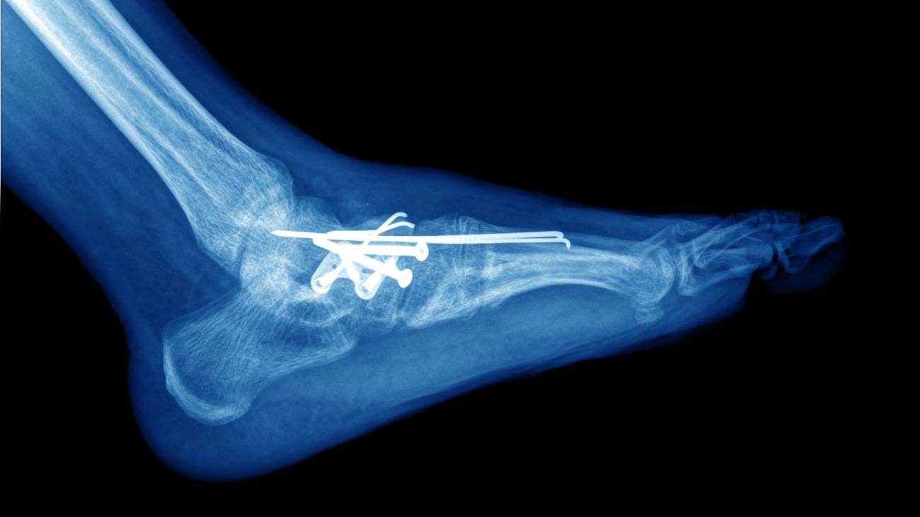
- Reduction: This is the first stage and involves manipulation of the bone fragments with the aim of returning them to their native position. Depending on the case, this is done by means of a closed reduction or without surgical opening of the fracture site, which is less aggressive, but sometimes does not allow an anatomically perfect reduction to be achieved. In this case, an open reduction or reduction with opening is required, which involves a surgical approach to the fracture site and, although more severe, allows the bone structure to be returned to its natural position.
- Stabilisation: Subsequently, in the second phase, the aim is to keep the reduction completely stable over time. For this, different methods can be used, which are established for each patient, depending on various factors relevant to the injury. Generally, however, stabilisation is achieved by plaster or traction (non-surgical means) or by pins, screws, plates or external fixators (methods involving an operation).
- Rehabilitation: Finally, the patient undergoes the rehabilitation stage from which it is hoped to restore function to the affected bone segment as efficiently and quickly as possible. To achieve this, a personalised physiotherapy plan is generally used to treat the consequences of the injury by promoting healing, treating soft tissue damage, reducing the effects of immobilisation and preventing future complications.
What methods of prevention to avoid broken bones are the most effective?

Although it is impossible to avoid pathological fractures due to certain conditions that naturally produce this type of injury, the truth is that people can implement different methods of care and prevention to avoid this type of contusions that generate a considerable symptomatological picture.
We advise you to practice the following simple and effective recommendations:
- Eat foods rich in calcium, collagen and vitamin D regularly: For, these substances are essential for preserving bone health and strengthening bone density.
- Engage in moderate physical activity: By practising sports, you can promote bone strengthening, tone muscles and improve your balance. This, as long as they are high-impact sports to promote bone structure (walking, running, dancing, lifting weights, etc.) and without overexerting the body.
- Adopt healthy lifestyle habits: Having a healthy and balanced diet, as well as avoiding the consumption of alcohol and tobacco, will allow you to avoid bone fractures. Harmful habits tend to reduce bone density, cause disorientation and slow reflexes (especially alcohol consumption).
- Wear the right footwear for your feet: It is also essential that people wear comfortable, low-heeled, rubber-soled, non-slip shoes to prevent the sudden falls that trigger fractures.
- Proper equipment: When you want to practice any sport, it is important that you use the right protective equipment to prevent injuries. Be it helmet, knee pads, elbow pads, ankle pads, etc.
- Prevent accidents: If you are in a hurry while driving or walking in the street, keep in mind that it is pertinent to slow down. Because, accidents are more likely to occur when people speed up (both their walking motion and their car).
- Avoid accidents in the home: In your home, make sure to keep the floor in a safe place to avoid accidents. To do this, it is appropriate to keep them free of clutter, remove carpets and loose cables, do not use slippery wax, leave furniture in its usual place, etc.
- Be careful with showers: In the bathrooms of your home, it is appropriate to have a non-slip rubber mat in the shower and to install grab bars on the bathroom walls (next to the shower, bathtub and toilet). This will reduce the risk of slipping and fractures.
- Keep your house clean: In the kitchen, it is recommended to clean up food spills as soon as they occur (or in any area of the house), as well as to use non-slip mats on the floor near the cooker and sink to prevent falls.
- Prepare your home: On stairs, to avoid sudden eventualities, you can choose to install sturdy handrails on both sides, mark the top and bottom step with bright tape and keep stairs well lit (with light switches at the top and bottom).
- In bedrooms, it is also a good idea to place light switches within reach of your bed (or a battery-operated torch, at the very least) and to get up slowly when lying or sitting on the bed. This will help prevent home accidents.
F.A.Q: Frequently asked questions
With regard to bone fractures, there are still many doubts on the part of people who are interested in preventing these injuries, and we will try to resolve them in the following sections:
References
- Müller, M. E., Nazarian, S., Koch, P., & Schatzker, J. (2012). The comprehensive classification of fractures of long bones. Springer Science & Business Media. https://books.google.es/books?hl=en&lr=&id=t0CgBQAAQBAJ
- Watson-Jones, R. (1962). Fractures and joint injuries. E. & S. Livingstone. http://117.239.25.194:7000/jspui/bitstream/123456789/1853/7/INDEX.pdf
- Hoppenfeld, S., & Murthy, V. L. (Eds.). (2000). Treatment and rehabilitation of fractures. Lippincott Williams & Wilkins. https://books.google.com/books?hl=en&lr=&id=bxhycwgJUtQC
- Kanis, J. A., Borgstrom, F., De Laet, C., Johansson, H., Johnell, O., Jonsson, B., ... & Khaltaev, N. (2005). Assessment of fracture risk. Osteoporosis international, 16, 581-589. https://link.springer.com/article/10.1007/s00198-004-1780-5
- BAILEY, M. (1982). Emergency! First aid for fractures. Nursing2021, 12(11), 72-81. https://journals.lww.com/nursing/Citation/1982/11000/EMERGENCY__FIRST_AID_FOR_FRACTURES.22.aspx
- Klotzbuecher, C. M., Ross, P. D., Landsman, P. B., Abbott III, T. A., & Berger, M. (2000). Patients with prior fractures have an increased risk of future fractures: a summary of the literature and statistical synthesis. Journal of bone and mineral research, 15(4), 721-739. https://asbmr.onlinelibrary.wiley.com/doi/full/10.1359/jbmr.2000.15.4.721
- Court-Brown, C. M., & Caesar, B. (2006). Epidemiology of adult fractures: a review. Injury, 37(8), 691-697. https://www.sciencedirect.com/science/article/abs/pii/S0020138306003238
- Singer, B. R., McLauchlan, G. J., Robinson, C. M., & Christie, J. (1998). Epidemiology of fractures in 15 000 adults: the influence of age and gender. The Journal of bone and joint surgery. British volume, 80(2), 243-248. https://online.boneandjoint.org.uk/doi/abs/10.1302/0301-620X.80B2.0800243
- Bischoff-Ferrari, H. A., Willett, W. C., Wong, J. B., Giovannucci, E., Dietrich, T., & Dawson-Hughes, B. (2005). Fracture prevention with vitamin D supplementation: a meta-analysis of randomized controlled trials. Jama, 293(18), 2257-2264. https://jamanetwork.com/journals/jama/article-abstract/200871
- Patel, D. S., Roth, M., & Kapil, N. (2011). Stress fractures: diagnosis, treatment, and prevention. American family physician, 83(1), 39-46. https://www.aafp.org/pubs/afp/issues/2011/0101/p39.html