- Definition: What is bursitis or inflammation of the bursae?
- What are the most common types of bursitis?
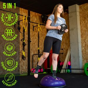
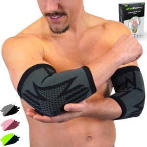
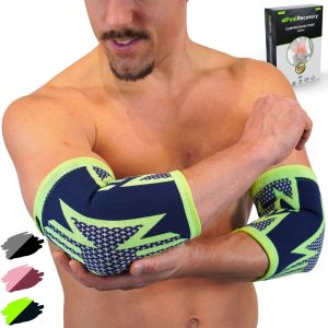
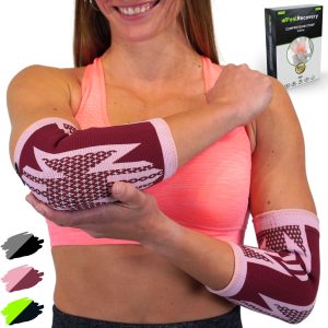
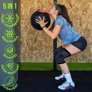
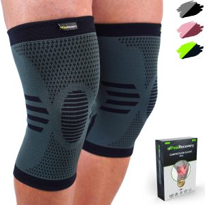
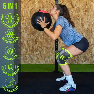
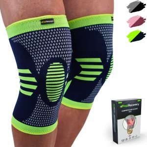
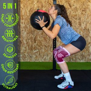
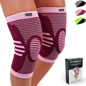
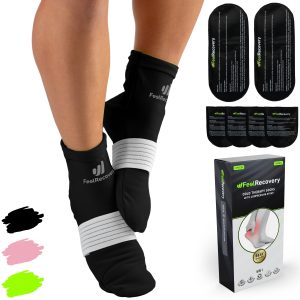
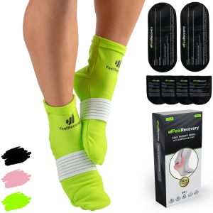
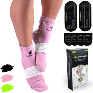
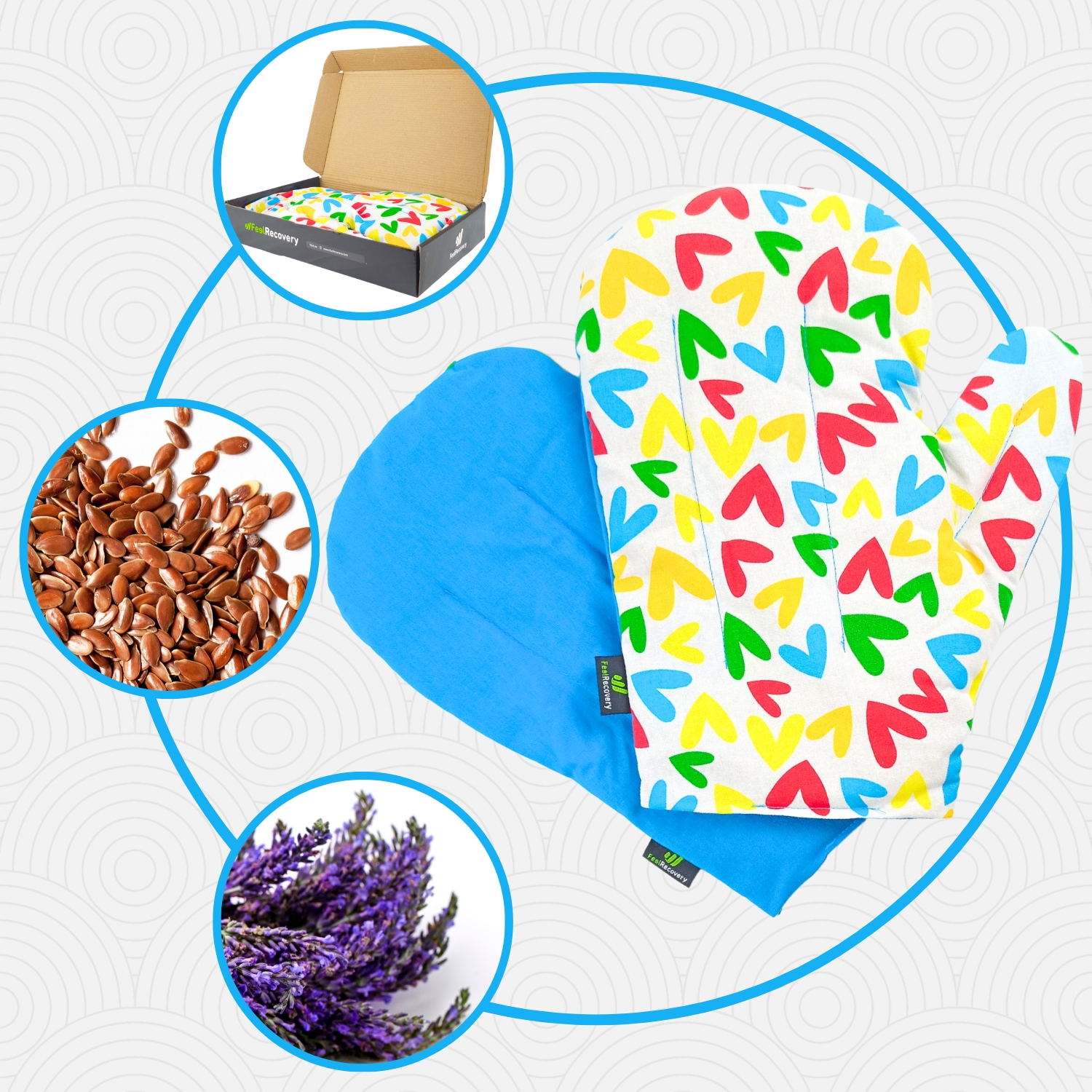
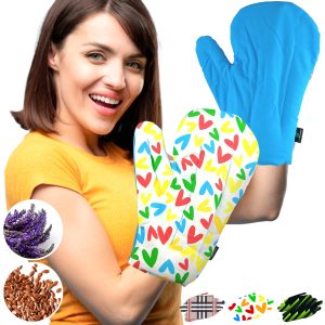
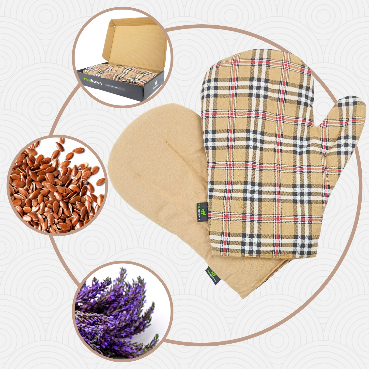
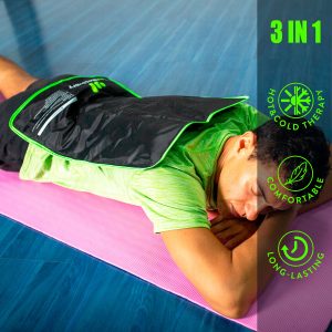
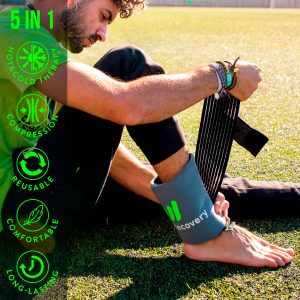
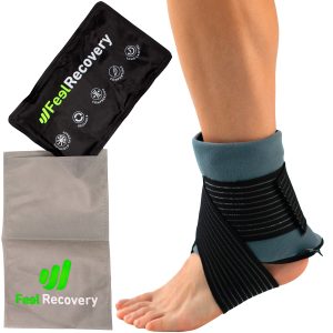
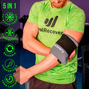
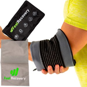
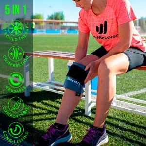
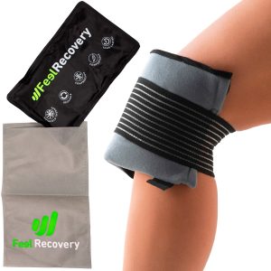
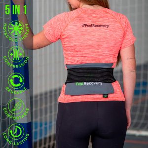
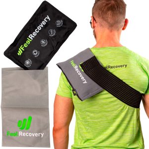
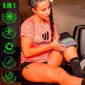
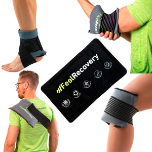
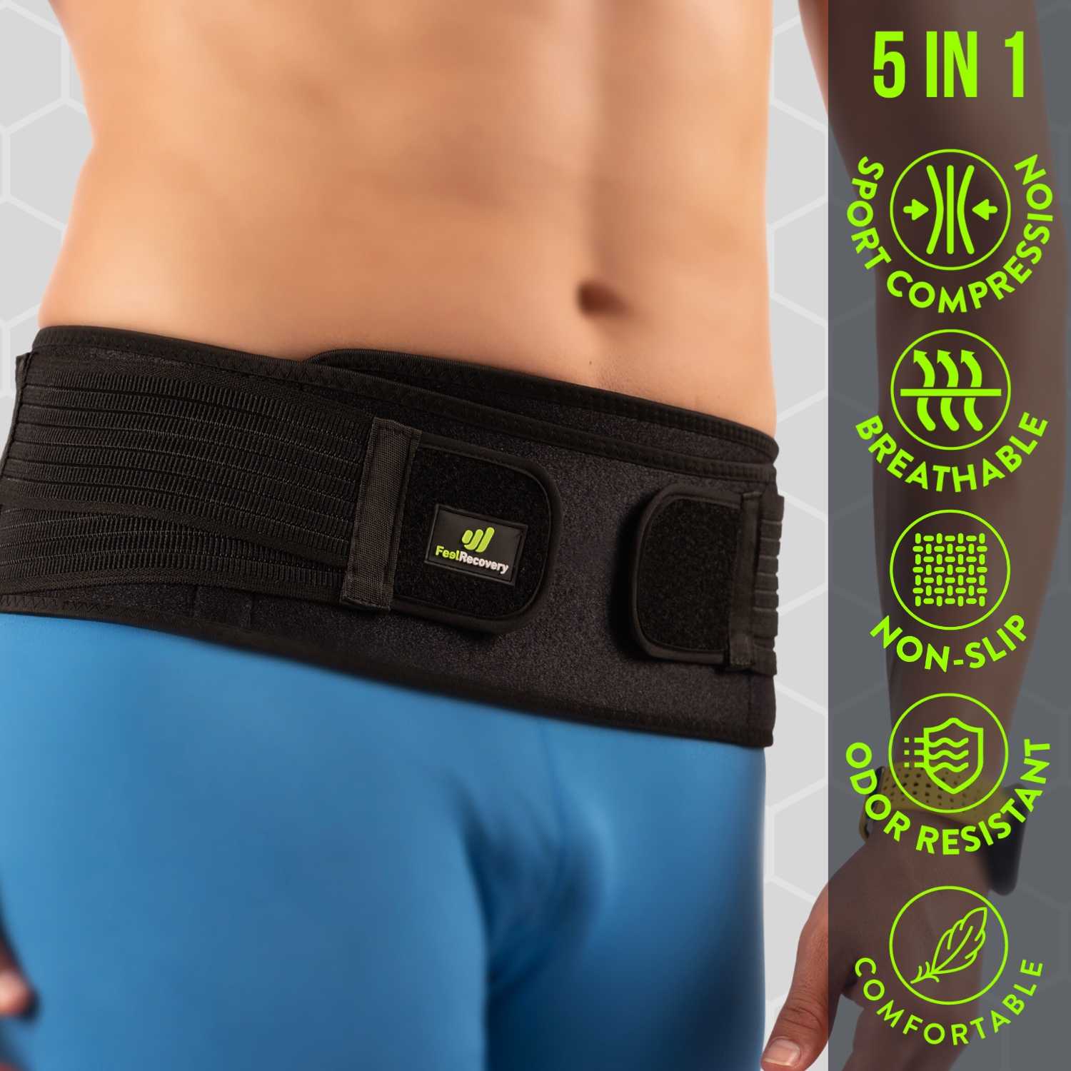
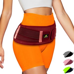
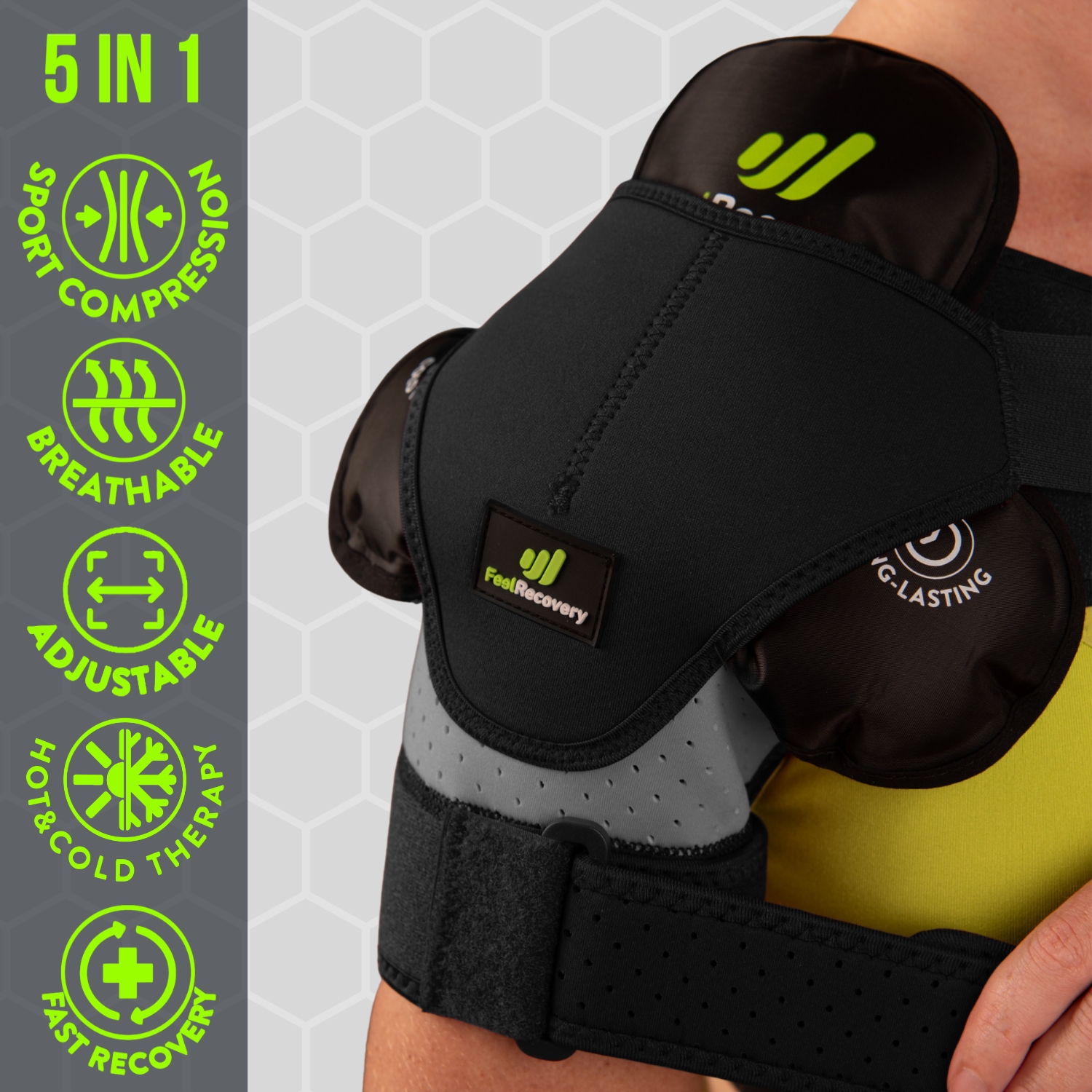
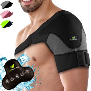
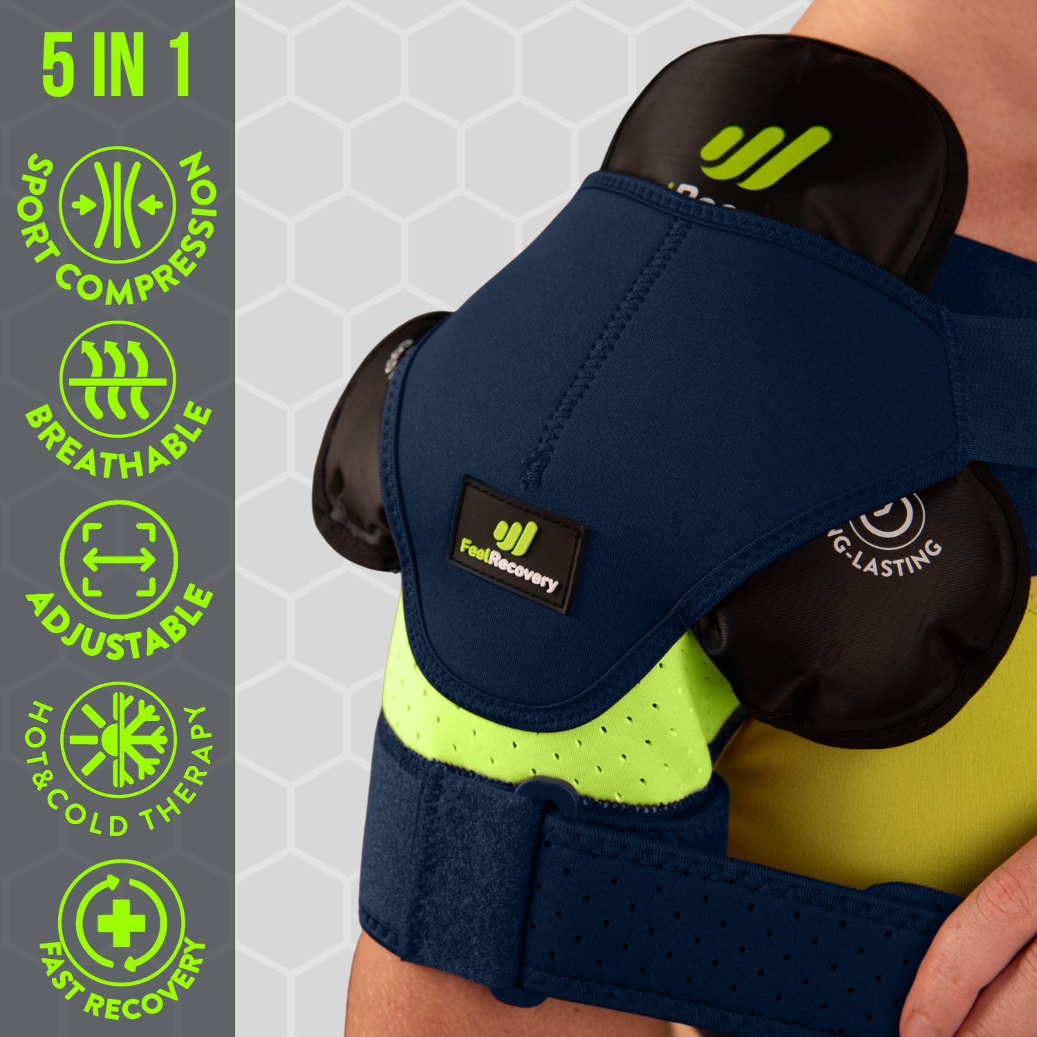
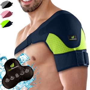
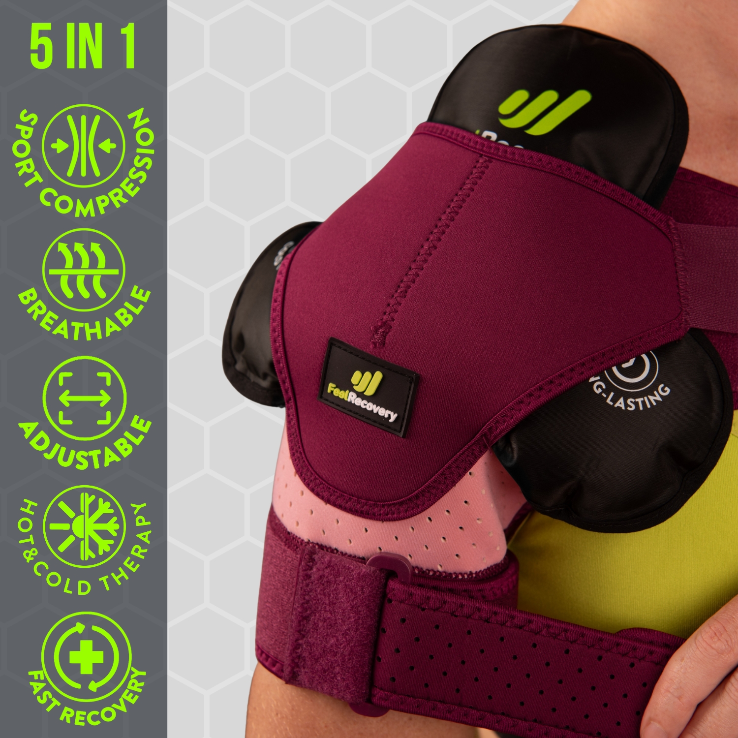
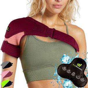
- Best bursitis products
- What are the causes and risk factors for bursitis?
- Main symptoms that warn us that we have bursitis
- What treatments are available to improve the symptoms of bursitis?
- What are the most effective prevention methods for bursitis?
- F.A.Q: Frequently asked questions
Inflammation of the synovial bursae is a pathology that occurs in the joints, which can cause pain and immobility. For this reason, you should know what bursitis is and what the most common types are. This information is explained in a simple way in the following paragraphs.
In addition, you can read about the factors that increase the likelihood of contracting bursitis, as well as the main symptoms that will warn you of the presence of the disease. Finally, you will find the current treatments and frequently asked questions about bursitis.
Definition: What is bursitis or inflammation of the bursae?
Bursitis is a pathology caused by inflammation in the synovial fluid sacs, which are located in the joints. These bursae, are specifically located between bones, tendons and muscles. Their function is to allow all parts to glide together so that joint movement can occur without direct friction between the bones.
However, if for some reason they become overloaded, injured or infected, the immune system produces an excess of synovial fluid, often making movement impossible or making it difficult to open the joint properly. This is when the visible signs of the disease, such as swelling or lumps, occur. It should be borne in mind that this situation can occur in different body regions such as elbows, knees, shoulders and hips among other joint areas.
What are the most common types of bursitis?

We will show you below the different types of bursitis that can occur in each joint. See below:
According to the types
According to the time and type of symptoms of the disease, bursitis can be classified as follows:
Acute bursitis: the characteristic of acute bursitis is its abrupt or sudden onset. Within a short time, inflamed and discoloured tissue is seen. The condition is brought on by injury, overuse, soft tissue pressure or infection. This diagnosis is possible if the skin has a high temperature in the affected area. It is also possible that this type of bursitis is caused by previous infections from a contaminating agent. This is more common in middle-aged male patients or those with gout.
Chronic bursitis: If a successive acute attack occurs in the same joint area, chronic bursitis is likely to be present. This type of bursitis can arise after treatment of the acute condition in which the necessary measures were not taken. A terrible mistake made by sufferers of this ailment is not to see a doctor in time. At this point, the problem can be perpetuated for weeks and months, resulting in small crunches of the bones when the joint is moved. From therapies to injections, it is the doctor who will decide the guidelines to follow to achieve remission.
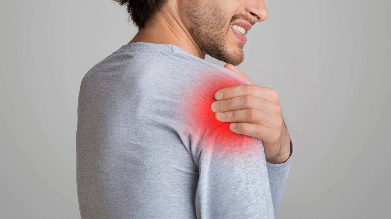
Subacromial bursitis: one of the most common, it disturbs one of the most axially mobile joints in the human body, the shoulder. In this ailment, the synovial bursa located between the acromion (lateral bone that acts as the natural bony stop of the scapula), the tendons and the trochlea is inflamed, affecting, in particular, the rotator cuff tendons. This causes pain and a feeling of heaviness; which, by the way, is likely to increase if the arm is rotated, extended or elevated. This bursitis most commonly occurs in people who are physically active and require shoulder movements, although it is also possible to find causes related to cartilage wear and tear, bone malformation or degenerative conditions.
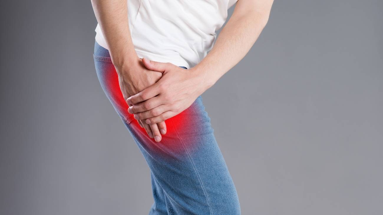
Ischial bursitis: is also called ischial or ischiogluteal bursitis because the tenderness or bone friction is felt in the hip and, in some cases, even in the back of the thigh. It can cause tingling and loss of sensation in the lower legs. The condition occurs in the ischial bursa, over the ischium (bone that forms part of the lower back part of the pelvis) and between the gluteus maximus. It is likely that the appearance of this disease is due to the fact that the person spends a lot of time sitting down or because of the practice of sports or work in which the hip movements are overdemanded and pressure is maintained for a long time on the ischium.
Trochanteric bursitis: this is another common variant of bursitis, also called trochanteritis. In this ailment, the pain intensifies in the external, lateral and more pronounced area of the hip and can affect more female patients. Its main symptom is an inflammation of the synovial fluid sacs located between the gluteal tendons and the trochanter, especially the superficial bursa that covers the greater trochanter to support the friction and tension of the joint. The likelihood of developing this disease increases in people who are sedentary, obese, have osteoarthritis or spurs. Also in those with flat feet or who have injured their hip after a fall.
According to the area
Bursitis can be classified according to where the affected bursa is located in the body. Keep in mind that hundreds of bursae can be found, so we will mention only those joints in which this disease appears most frequently.
Have a look:
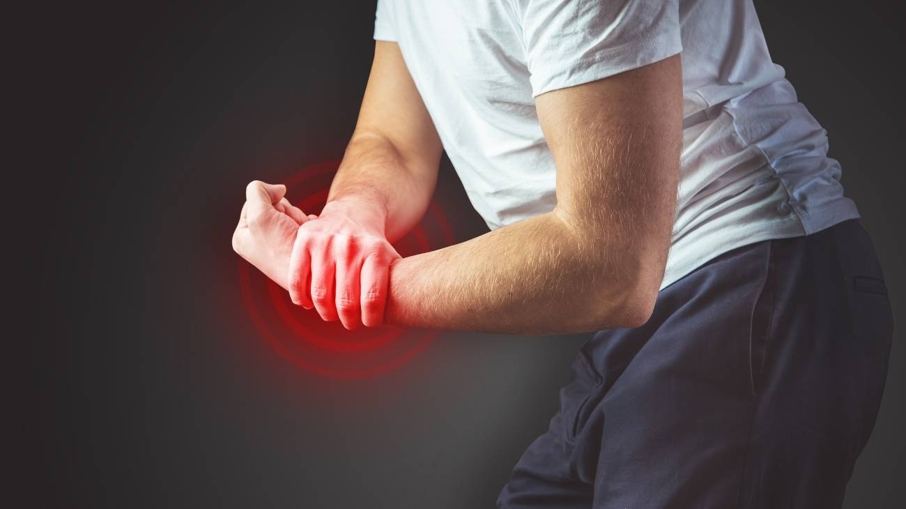
Bursitis of the hands and wrists: this area has two main bursae that do individual work, the ulnar and the radial. These cover every tendon from the hand to the wrist. The first one works for the tendons that go to the little, index, ring and middle fingers, while the second one helps the movements of the thumb. Wrist bursitis can occur as a result of a blow, fall, joint wear and tear, infection (horseshoe abscess) or tertiary complications. In this form, the pain is worse when writing, lifting a heavy object or simply bending the joint. In a few severe cases the specialist will choose to remove the bursa. Swelling usually subsides by avoiding joint stress.
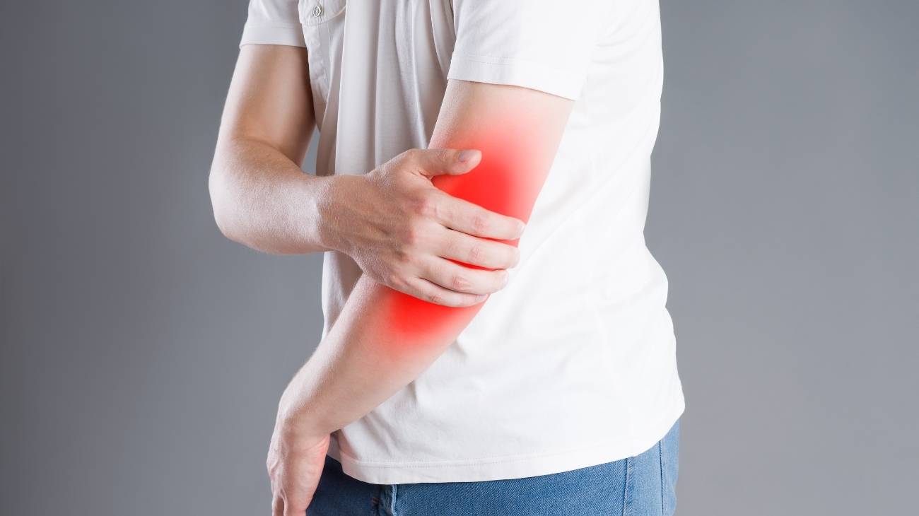
Elbow bursitis: in this mobile area there is little soft tissue to cushion friction. As such, the bursae do much of the work of bearing the pressure of movement, it is common for them to become inflamed, especially in male patients. The condition is also called olecranial bursitis because the bursa is located at the back of the elbow, at the olecranon. Once the bursa fills with fluid, it is possible to see bulges and there may be key triggers for this condition. For example, people who rest their elbow on the table for long periods of time, rheumatic diseases and infectious cases, which occur less frequently.
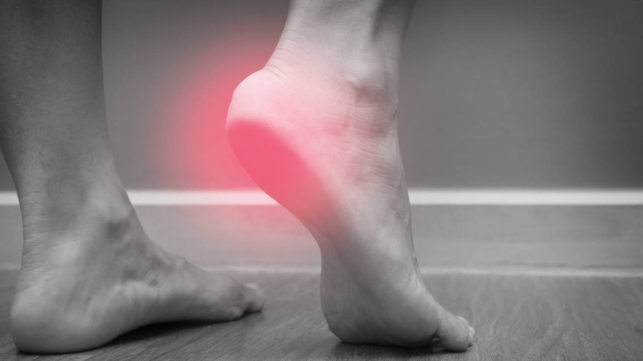
Achilles tendon bursitis: this is one of the most common due to the complexity of the anatomy of the foot and the number of joints. This disease is linked to the Achilles tendon and is generated directly in the inner rear part of the feet. Two synovial bursae of different sizes are located there, the smallest being the retrocalcaneal bursa and the largest being the subcutaneous calcaneal bursa. They owe their names to the fact that they are located between the Achilles tendon, the calcaneal bone and the skin. It is usually the more predominantly posterior one that is affected because it withstands the pressure between the skin and the tendon that it suffers as a result of improper footwear. A large percentage of patients improve simply with therapeutic treatment and rest.
Shoulder bursitis: in this area of the body, the synovial pads have a minimal amount of fluid to protect the friction between bones and other tissues, so this type of bursitis is also common. This ailment is caused by a disorder of the subdeltoid or subacromial bursa, located under the deltoid muscle and the acromion, which is responsible for causing correct sliding in the joint. There is a higher incidence of this disease in cases of sportsmen or manual workers as well as in diseases with degenerative processes such as tendinitis. Shoulder impingement is another factor to be taken into account for diagnosis.
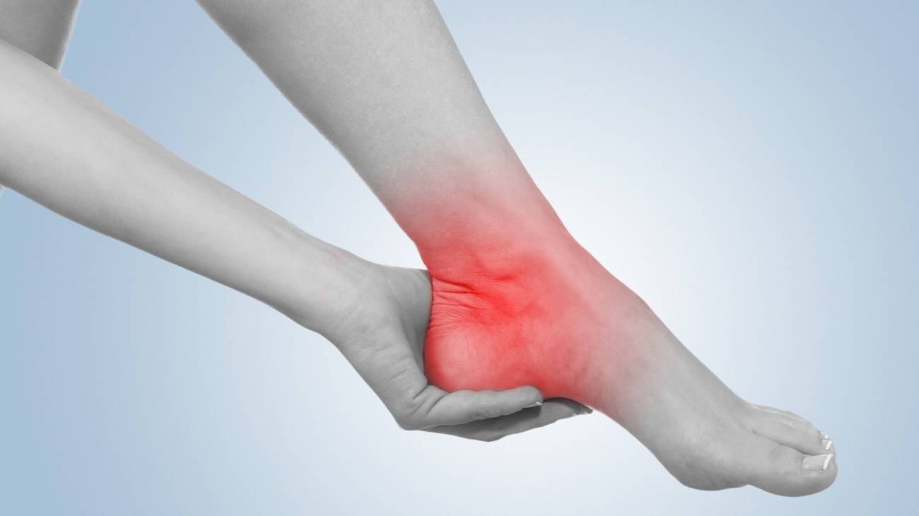
Bursitis of the ankle: in this part of the body there are 3 bones that fit into the talus and between the talus, the lower heel bone (the calcaneus) and the Achilles tendon it is possible to find a bursa. This synovial bursa, called the retrocalcaneus, is responsible for facilitating the ankle joint. When its volume increases, bursa trauma of the ankle, also called Albert's disease, occurs. This can be caused by overweight, sprains, repetitive movements, poor bone formation and hereditary factors. In very rare cases, Haglund's syndrome (enlargement of the bony parts of the foot) also leads to this pathology.
Hip bursitis: when the hip is overloaded, the bursa covering the trochanter bone is affected, which is why trochanteritis is the most frequently mentioned. However, the ischial bursa (near the ischium bone) is also part of the inflammatory pathologies of this area of the body. Patients with these conditions describe a sharp, intense pain that makes it difficult to sleep and are usually female and middle-aged. Those who have had hip surgery are also potentially at risk. Of all the modalities, it is one of the most enduring that is prone to become chronic if not treated in time.
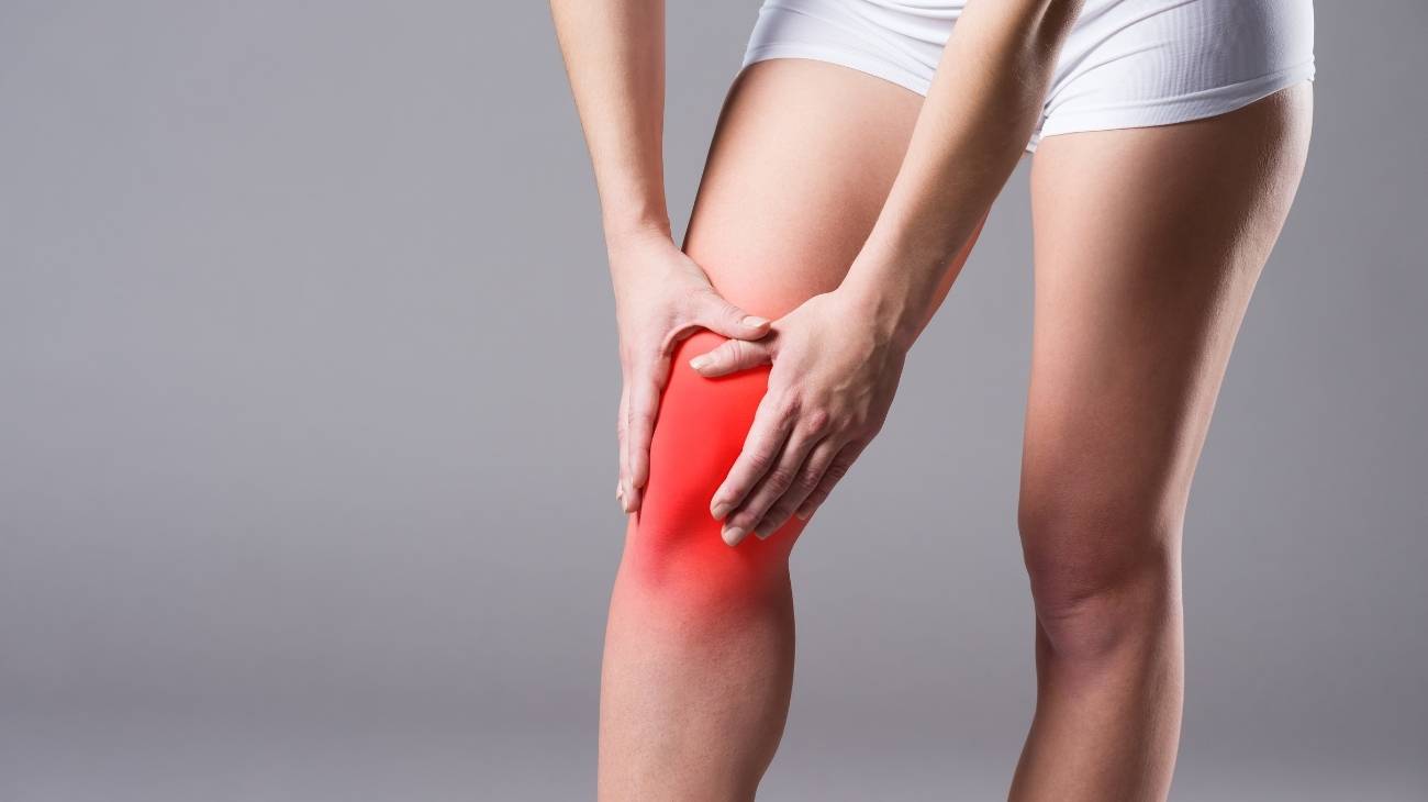
Knee bursitis: Up to eleven bursae can be found in the knee. Once one of these is disturbed, the mobility of the area is limited, causing intense pain and, in some cases, swelling. Among the most common is patellar bursitis, which affects the superficial and deep bursa, and is common in people who spend a lot of time on their knees. It is also common to find patients with inflammation of the bursa on the inside of the knee, known as Pes Anserine, which can be medial, if it is the semimembranosus bursa, or prepatellar, if it is in the anterior area, and is prone to haemorrhage and bruising. The doctor is likely to recommend cold therapy, along with rest, to reduce swelling.
Best bursitis products
Bestseller
-

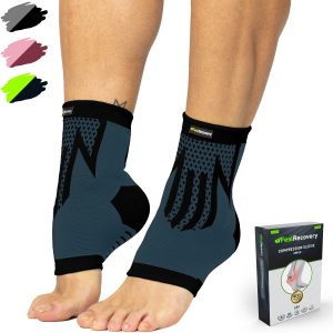
2 Ankle Compression Sleeve (Black/Gray)
$19.95 -
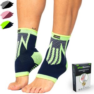
2 Ankle Compression Sleeve (Green/Navy)
$19.95 -
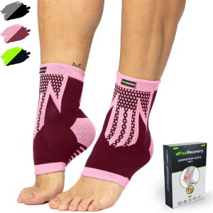
2 Ankle Compression Sleeve (Pink/Bordeaux)
$19.95 -
2 Elbow Compression Sleeve (Black/Gray)
$19.95 -
2 Elbow Compression Sleeve (Green/Navy)
$19.95 -
2 Elbow Compression Sleeve (Pink/Bordeaux)
$19.95 -
2 Knee Compression Sleeve (Black/Gray)
$19.95 -
2 Knee Compression Sleeve (Green/Navy)
$19.95 -
2 Knee Compression Sleeve (Pink/Bordeaux)
$19.95 -

Ice Pack for Foot - Cold Therapy Socks (Black)
$24.95 -
Ice Pack for Foot - Cold Therapy Socks (Green)
$24.95 -
Ice Pack for Foot - Cold Therapy Socks (Pink)
$24.95 -
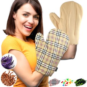
Microwave Arthritis Gloves (2 Mittens) (Hearts)
$29.95 -
Microwave Arthritis Gloves (2 Mittens) (Oxford)
$29.95 -
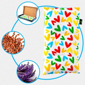
Microwaveable Heating Pad for Pain Relief (Hearts)
$19.95 -
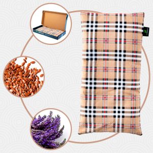
Microwaveable Heating Pad for Pain Relief (Oxford)
$19.95 -
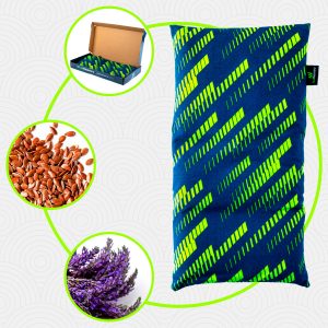
Microwaveable Heating Pad for Pain Relief (Sport)
$19.95 -
Reusable Extra Large Ice Packs for Back & Legs Pain
$29.95 -
Reusable Gel Ice Packs for Ankle & Foot with Compression Band
$19.95 -
Reusable Gel Ice Packs for Elbow & Arms with Compression Band
$19.95 -
Reusable Gel Ice Packs for Knees with Compression Band
$19.95 -
Reusable Gel Ice Packs for Shoulder & Back with Compression Band
$19.95 -
Reusable Gel Ice Packs with Compression Band for Sports Injury
$19.95 -
Reusable Ice Pack for Neck & Shoulders Injuries
$24.95 -
Sacroiliac Support Belt (Black)
$24.95 -
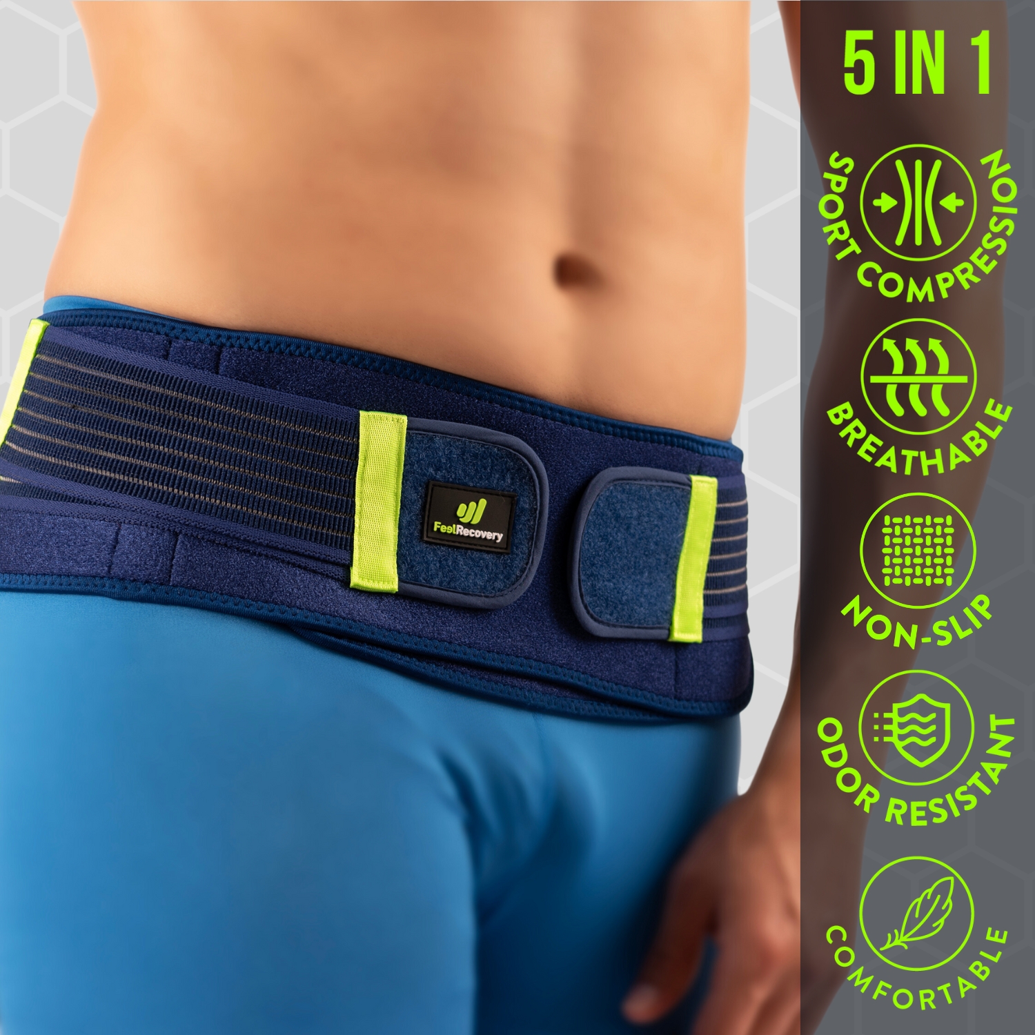
Sacroiliac Support Belt (Green)
$24.95 -
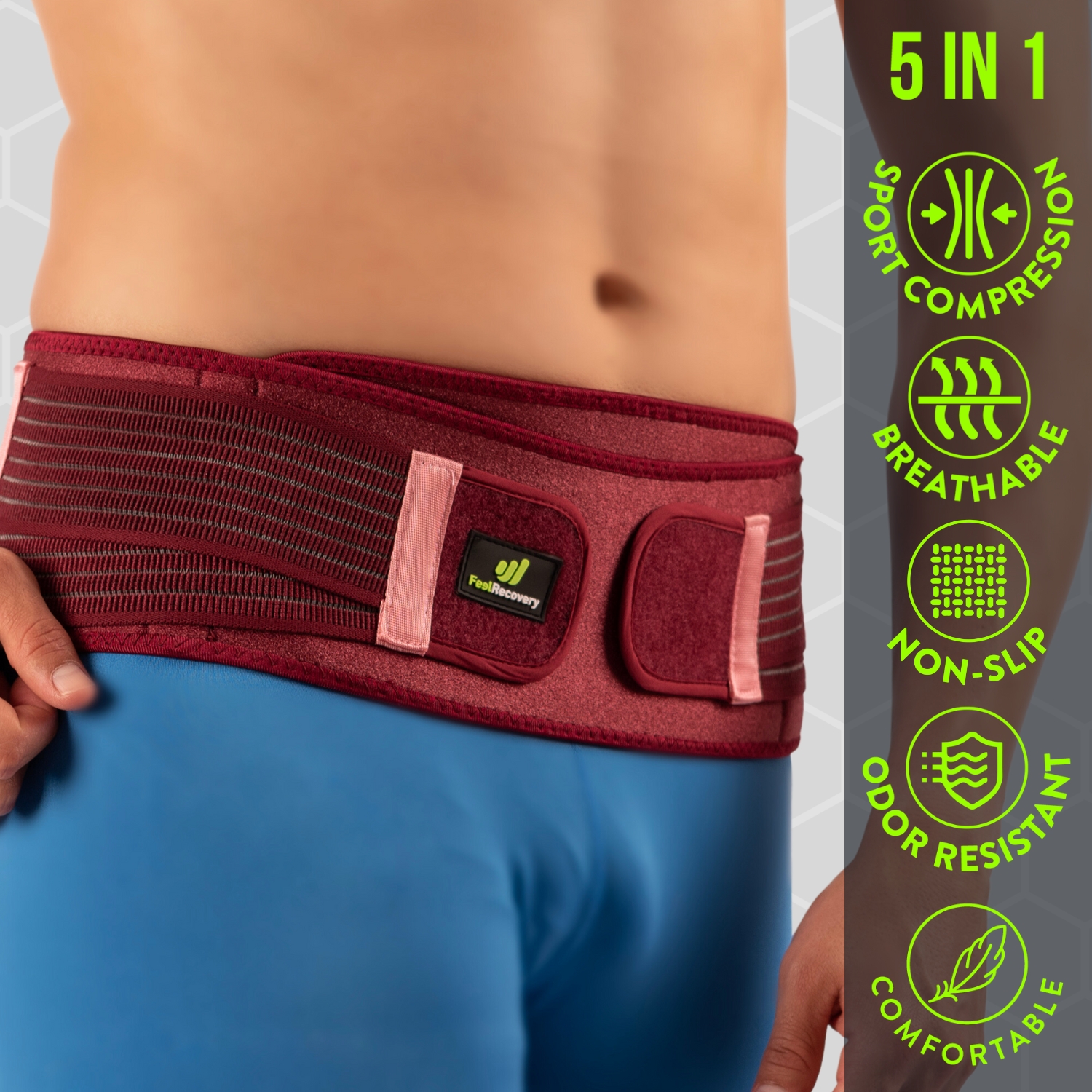
Sacroiliac Support Belt (Pink)
$24.95 -
Shoulder Support Brace (Black)
$24.95 -
Shoulder Support Brace (Green)
$24.95 -
Shoulder Support Brace (Pink)
$24.95 -
Wrist Brace (Black/Gray)
$19.95 -
Wrist Brace (Green/Navy)
$19.95 -
Wrist Brace (Pink/Bordeaux)
$19.95


What are the causes and risk factors for bursitis?
One aspect that will help to improve the symptomatology of bursitis is to determine the cause or the factor that caused it, so that a recurrence can be avoided in the near future. It may even help to work on the area or to establish the best treatment.
Keep in mind that it is these circumstances that increase the likelihood of contracting the disease. These factors are discussed below:
- Overloading: the strain on the joints every time weights are lifted can lead to crushing of the synovial cavity, which can lead to inflammation of a bursa.
- Repetitive mechanical work due to a certain activity or profession: while it is true that overloading places stress on the joint, this is not the only cause of stress. Constant and repeated movements can affect the synovial fluid within the bursa, leading to inflammation and pain in the area.
- Trauma: poorly healed fractures or injuries to the joint can affect the joint capsule. There are several factors for this, but the most recurrent are the septic aspects and the osteophytes that may remain after the trauma has healed. This causes the joint to malfunction generating inflammation in the bursa.
- Injuries: a separate point from trauma are blows to the synovial bursa. This can stimulate the onset of the disease because the joint is seriously affected in its functioning.
- Sudden movements: violent actions that are carried out with the joint overdemand it, stimulating the synovial bursa to not work with enough space, which can create inflation in the bursae.
- Obesity: people with excess body weight have an additional risk factor for the development of bursitis. This is due to the constant overloading of the joints when walking, standing or bending down, causing the bursa containing the articular cartilage to be affected in space, which causes a malfunction of the joint.
- Suffering from rheumatoid arthritis: this chronic, degenerative disease has its consequences when it deforms the joints. One of them is the appearance of inflation in the bursae.
- Suffering from diabetes: having an excess of glucose in the blood due to a lack of insulin can lead to fatigue and musculoskeletal changes. This leads to stiffness in the muscles, causing poor performance of the joint and thus the bursae.
- Gout disease, osteoporosis and fibromyalgia: are some of the diseases that cause inflammatory changes in the synovial bursae. These diseases cause stiffness and weakness in the joint areas.
- Age: in this risk factor it is necessary to distinguish by type of bursitis. Men around 50 years of age are more likely to get inflammation in the superficial bursae, while women of 55 years of age are more likely to suffer from deep bursitis.
Main symptoms that warn us that we have bursitis

Symptomatology may vary according to the region affected. For example, in the knee, more swelling or bruising is seen, while in the hip, severe pain is one of the main indicators. However, there are common patterns that are obvious signs of bursitis.
If you feel any of these signs of the disease, it is best to see a doctor as a matter of urgency:
- Feeling of joint heaviness or stiffness: this may be accompanied by pain in the joints, which are characteristic symptoms of the presence of bursitis. This is caused by various factors, the most common being the accumulation of synovial fluid and tension in tendons and muscles in the affected area.
- Inability to perform everyday movements or exercises: it is possible to become aware of the disease when the patient finds it difficult to perform everyday tasks. The stiffness is usually caused by the involuntary tension that the person exerts to reduce the pain.
- Swelling: the large amount of synovial fluid in the joint capsule can cause the cavity to swell, thus causing an obvious change in the volume of the joint.
- Abnormal lumps: it is possible to find the presence of nodules or superficial lumps in these types of ailments. In these cases, a doctor should be consulted as soon as possible to rule out the presence of other diseases and to treat the bursitis.
- Reddening of the joint area: the lack of blood supply to the dilator vessels means that they do not work properly and the blood does not circulate properly, which causes the muscles and tendons to become unusually red.
- Skin sensitivity in the affected region: the skin is affected by bursitis, so the nerves in the affected area become more sensitive to pain, causing any friction to generate pain.
- Fever: this symptom may be accompanied in some patients by sudden tremors and night sweats.
What treatments are available to improve the symptoms of bursitis?
Whatever the cause, it is necessary to see a doctor to diagnose the condition. The doctor is likely to ask questions, carry out a physical and radiological examination to establish the presence of bursitis. In the first instance, conservative treatment is given if the cause is not serious or infectious, so that the inflammation in the synovial bursa usually improves within a few days.
If the results show a more severe infection, any of the following methods can be applied:
Alternative and complementary therapies
The following therapies can be applied to patients with bursitis:
- Heat and cold therapy: in these cases a treatment is applied that includes heat to reduce muscle stiffness and cold to deflate the affected area, among other effects. In this way it is possible to apply 3 to 4 times a day until the symptoms gradually subside, but it is important to bear in mind that this technique should not exceed 20 minutes of application per session. Different elements are used to transmit the desired temperature, from bags with hot or cold water, localised baths (depending on the joint) and compresses with special gels. Ointments or ointments should never be used in this treatment, as these creams could severely affect the skin and cause burns.
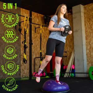
- Compression therapy: by placing different elastic bandages, compression garments or braces supports designed for certain regions, blood circulation is improved and the joint is immobilised, as well as compressing the venous walls in a controlled manner. It is widely used in knee conditions to reduce chronic inflammation, heaviness and swelling. It is necessary to consult a doctor before using these products because they may induce injury by compressing the synovial sacs.
- Massage therapy: Some physiotherapists often recommend guided self-massage, e.g. for trochanteric bursitis. These are massages of stretching and gentle movements where it is advised to avoid directly massaging the area close to the bone. In addition to this, it is possible to use a professional to use different techniques to reduce stiffness and inflation. The ultimate goal is for the patient to feel general relief.
- Acupressure therapy: with this Chinese technique, pressure is applied to different parts of the body to reduce muscle tension, improve blood flow and increase the patient's sense of wellbeing. This treatment is performed using only the palms and fingers of the hand to apply pressure to strategic areas.
- Thermotherapy: the use of heat is productive in causing blood vessels to dilate. This considerably improves the reduction of joint stiffness by causing a greater opening of the capsule containing the bursa. In this way it is possible to reduce the inflammation within a few days. As with heat and cold therapy, sessions should not exceed 15 to 20 minutes and various elements can be used to radiate heat. For example, special gels, electric blankets and hot water bottles.
- Natural remedies using plants: There are hundreds of natural plant-based alternatives to relieve pain and swelling. Chilli, or chilli pepper, is part of ointments that reduce pain due to its capsaicin (a natural analgesic). Green tea can also be found in these therapies as a good anti-inflammatory and antioxidant. Flaxseed oil contains active compounds beneficial for joint lubrication, as do turmeric, ginger and apple cider vinegar. These natural plants should be seen as a beneficial adjunct to, but not a replacement for, medical treatment.
- Healthy lifestyle habits: the first thing any doctor will recommend is to replace the practices that have led to the person's overload. For example, regulating sports activities or mechanical work. For this, occupational therapy is a good joint measure. He or she will also recommend establishing a diet with the help of a nutritionist if you are overweight. This is where certain natural food supplements can come into play to help strengthen muscles and so on. Of course, these should be regulated by health specialists before use.
Dietary supplements
When patients need to incorporate substances into the body that are not present in the body, it is necessary to resort to additives. Among the most common are:
- Glucosamine: also known as glucosamine sulphate. It works to treat pain caused by osteoarthritis or other soft tissue disorders, although it is not recommended for use in people with asthma. It can also influence blood sugar levels.
- Fish oil: A formula promoted by some naturopaths to improve the signs of inflammation is fish oil. Because it contains special fatty acids such as Omega 6 and Omega 3, it is an anti-inflammatory remedy. All supplements from enzymes to natural oils such as soybean or fish oil should be consulted with your GP before use.
- Other supplements: magnesium, selenium, hyaluronic acid and chondroitin sulphate are some components that can also be found in powders, syrups, tablets and food liquids.
Physiotherapy treatments
In terms of application, physiotherapy can be direct, underwater or mixed. This will be decided by the therapist, depending on the area and the patient's aggravation, with underwater being a widely used strategy to eliminate pain. Other times it is used as a means of activating a cortisone ointment.
Physiotherapy includes:
- Controlled exercise routines: The visit to the physiotherapist will always be aimed at improving flexibility, strengthening the joint structure and elasticity of muscles and tendons. According to the patient's age, condition and other factors, an exercise routine is established. A combination of stretching and gentle repetitions (with or without the help of balls and dumbbells, among other elements).
- Muscle massage: In rare situations physiotherapists use light muscle massage. These are intended to reduce pain due to tension. However, in almost all cases they tend not to opt for this as a first choice. More stimulation would only worsen the situation of an already sensitive bursa due to overload.
- Ultrasound: these devices make use of vibratory energy from 3 MHz to 20 GHz, with the latter achieving the greatest penetration into the internal tissues. Pulsed ultrasound is used for acute inflammation. In contrast to the use of heat, it promotes a particular improvement of inflammation and pain.
- Hydrotherapy: This technique is used to gradually facilitate mobility. With the help of water, the joints can be moved more easily and without pain, as the earth's gravity is avoided. It has been proven that this therapeutic treatment often improves breathing and the cardiovascular system.
- Laser: Symptoms can be alleviated by means of biostimulation at a deep level. In spite of being a relatively new method, favourable results have been demonstrated and it can reduce swelling, pain sensations and even avoid bone complications.
- Cryomassage: Unlike the simple therapy of cold compresses, cryomassage adds ice massage as a restorative means. Using ice or another cold element raises the temperature in order to reduce swelling. Its application is slow, gentle and maintained so that the tissues and blood vessels improve.
- TENS: this is one of the most commonly used types of electrotherapy for bursitis, together with pulsed US. This method uses transcutaneous nerve stimulation which must be regulated by a physiotherapist in terms of duration and intensity.
Medicines
Before starting this treatment, it should be taken into account that self-medication can lead to future problems and complications for the patient, so it is never advisable to take medication without first visiting a doctor.
The pharmacological therapy for the treatment of bursitis is based on:
- Antibiotics: depending on the microorganism that has generated the abnormality in the bursae, the doctor will prescribe one or another type of antibiotic. They must be taken according to the indications prescribed in the prescription. The most commonly prescribed are Clarythromycin, Erythromycin and Flucloxacillin. With their help the causative bacteria are eliminated and prevented from reproducing.
- Anti-inflammatory drugs: These drugs that doctors may recommend include ibuprofen, ketoprofen, aspirin and naproxen. While some are available over-the-counter in pharmacies, self-medication is not advised. Nonsteroidal anti-inflammatory drugs are intended to reduce inflammation and the resulting pain. Because of this, they are almost never maintained after remission of the disease.
- Corticosteroids: these are a last resort when all other methods fail to provide improvement. They are injected directly into the soft tissues and not in pill form, as they are synthetic replicas of cortisol. This is a hormone that the body produces naturally to shrink inflamed tissue.
Surgery and fluid removal
This type of treatment is applied in cases where the disease has advanced significantly, the most common surgical techniques are:
- Fluid aspiration: the specialist may use this therapy when infection is suspected. It consists of inserting a needle and draining with a syringe to reduce the excess fluid, which reduces the pressure in the area to reduce inflammation. Local anaesthesia is used in the process and is most commonly applied to the knee, elbow and shoulder. However, short-term deep aspiration can cause pain or swelling, so it is considered a more invasive treatment than therapies.
- Bursectomy: is a surgery where the bursae are removed. Such a procedure is only limited to severe or high-risk situations where the patient's condition warrants it. Before proceeding, all other conservative treatments are usually applied. It lasts about 20 to 30 minutes depending on the location of the bags. General, spinal or local anaesthesia is used depending on the case.
What are the most effective prevention methods for bursitis?

Although there are situations beyond the patient's control, habits make a difference to joint health. This is especially true if you have had an acute case and can prevent it from progressing to a chronic case.
If you want to prevent bursitis, pay attention to the following list of the most effective prevention methods:
- Warm up before repetitive activities: This will help the muscles and tendons in the joint area to stretch slowly, which will be better able to withstand sudden movements and weight bearing.
- Use a gradual exercise routine according to your physical condition: Remember that a sedentary lifestyle is one of the most common causes that occur in patients with bursitis, so playing sports will help your joints work properly. This will help to strengthen the muscles surrounding the joint area.
- Maintain a balanced diet: Eating a healthy diet and avoiding foods high in uric acid will help prevent microcrystals in the joint areas, which will cause overbones.
- Wear protection when playing different sports: Especially in sports where the joints are overtaxed and can be subjected to heavy blows or excessive loads to avoid the risk of trauma.
- Use cushioned pads for joints in contact with hard surfaces: If you need to walk for long periods of time in areas where the floor is hard, it is advisable to use accessories in your footwear to cushion your steps.
- Maintain proper posture: Healthy lifestyle habits include not only exercise and healthy eating, but also re-educating the patient to adopt beneficial movements. This includes learning how to sit, climb up or down stairs and lift heavy objects from the floor, among other things.
- Implement rest times between repetitive tasks: It is important to rest the joint when working or playing sports. This will help prevent wear and tear on the cartilage and keep the synovial fluid at optimal levels.
- Interrupt movements if you have noticed that activities are causing pain: If for any reason, the actions you are carrying out complicate the functioning of the joint causing pain in the form of a pinch, it is advisable to discontinue with the routine and visit the doctor.
- Stop smoking: Smoking is a very common risk factor for people suffering from bursitis. For this reason, it is advisable to stop smoking as well as drinking alcohol.
F.A.Q: Frequently asked questions
Here are the most common questions people ask about bursitis:
References
- McAfee, J. H., & Smith, D. L. (1988). Olecranon and prepatellar bursitis. Diagnosis and treatment. Western Journal of Medicine, 149(5), 607. https://www.ncbi.nlm.nih.gov/pmc/articles/PMC1026560/
- Reilly, D., & Kamineni, S. (2016). Olecranon bursitis. Journal of shoulder and elbow surgery, 25(1), 158-167. https://www.sciencedirect.com/science/article/abs/pii/S1058274615004693
- Khodaee, M. (2017). Common superficial bursitis. American family physician, 95(4), 224-231. https://www.aafp.org/pubs/afp/issues/2017/0215/p224.html
- Rasmussen, K. J. E., & Fanø, N. (1985). Trochanteric bursitis: treatment by corticosteroid injection. Scandinavian journal of rheumatology, 14(4), 417-420. https://www.tandfonline.com/doi/abs/10.3109/03009748509102047
- Sayegh, E. T., & Strauch, R. J. (2014). Treatment of olecranon bursitis: a systematic review. Archives of orthopaedic and trauma surgery, 134, 1517-1536. https://link.springer.com/article/10.1007/s00402-014-2088-3
- Lustenberger, D. P., Ng, V. Y., Best, T. M., & Ellis, T. J. (2011). Efficacy of treatment of trochanteric bursitis: a systematic review. Clinical journal of sport medicine: official journal of the Canadian Academy of Sport Medicine, 21(5), 447. https://www.ncbi.nlm.nih.gov/pmc/articles/PMC3689218/
- Baumbach, S. F., Lobo, C. M., Badyine, I., Mutschler, W., & Kanz, K. G. (2014). Prepatellar and olecranon bursitis: literature review and development of a treatment algorithm. Archives of orthopaedic and trauma surgery, 134, 359-370. https://link.springer.com/article/10.1007/s00402-013-1882-7
- Aaron, D. L., Patel, A., Kayiaros, S., & Calfee, R. (2011). Four common types of bursitis: diagnosis and management. JAAOS-Journal of the American Academy of Orthopaedic Surgeons, 19(6), 359-367. https://journals.lww.com/jaaos/Abstract/2011/06000/Four_Common_Types_of_Bursitis__Diagnosis_and.6.aspx
- Shbeeb, M. I., & Matteson, E. L. (1996, June). Trochanteric bursitis (greater trochanter pain syndrome). In Mayo Clinic Proceedings (Vol. 71, No. 6, pp. 565-569). Elsevier. https://www.sciencedirect.com/science/article/abs/pii/S002561961164113X
- Alvarez-Nemegyei, J., & Canoso, J. J. (2004). Evidence-based soft tissue rheumatology IV: anserine bursitis. JCR: Journal of Clinical Rheumatology, 10(4), 205-206. https://journals.lww.com/jclinrheum/Abstract/2004/08000/Evidence_Based_Soft_Tissue_Rheumatology_IV_.7.aspx